- Clone
- Poly6330 (See other available formats)
- Regulatory Status
- RUO
- Other Names
- E3 ubiquitin-protein ligase Cbl, Proto-oncogene c-Cbl
- Isotype
- Rabbit Polyclonal IgG

-

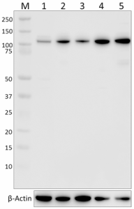
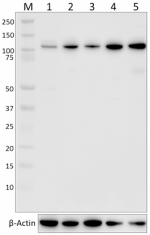
Total cell lysates (15 µg protein) from HepG2 (Lane 1), HeLa (Lane 2), Raji (Lane 3), HEL (Lane 4), or RAW264.7 (Lane 5) cells were resolved using 4-12% Bis-Tris gel electrophoresis, transferred to nitrocellulose, and probed with purified anti-Cbl antibody (Poly6330) at a 1:1000 dilution. Proteins were visualized by chemiluminescence detection using HRP Donkey anti-rabbit-IgG (Cat. No. 406401) at a 1:3000 dilution. Equal protein loading was confirmed using Direct-Blot™ HRP anti-β-actin antibody (Cat. No. 643807) at a 1:5000 dilution (lower). Lane M: MW ladder. Cell lysates were loaded in order of increasing CBL mRNA expression levels (source: Human Protein Atlas).
| Cat # | Size | Price | Quantity Check Availability | ||
|---|---|---|---|---|---|
| 633001 | 125 µL | $112.00 | |||
The proto-oncogene c-Cbl was initially identified as the cellular homologue of v-Cbl oncogene that induces pre-B cell lymphomas and myeloid leukemias in mice. The Cbl family comprises three mammalian proteins, c-Cbl, Cbl-b, and Cbl-3; among these, c-Cbl and Cbl-b are expressed in hemopoietic cells, whereas Cbl-3 is expressed only in epithelial tissues. Cbl proteins can function as E3 ubiquitin (Ub) ligases, and in more recent studies Cbl has been implicated in the negative regulation of various RTKs.
Product Details
- Verified Reactivity
- Human, Mouse, Rat
- Antibody Type
- Polyclonal
- Host Species
- Rabbit
- Immunogen
- peptide mapping to the carboxy terminal domain of human Cbl
- Formulation
- This antibody is provided in phosphate-buffered solution, pH 7.2, containing 0.09% sodium azide and 0.2% gelatin.
- Preparation
- The antibody was purified by affinity chromatography.
- Concentration
- Lot-specific (to obtain lot-specific concentration and expiration, please enter the lot number in our Certificate of Analysis online tool.)
- Storage & Handling
- The antibody solution should be stored undiluted between 2°C and 8°C. Do not freeze.
- Application
-
WB - Quality tested
- Recommended Usage
-
Each lot of this antibody is quality control tested by Western blotting. Western blotting suggested working dilution(s): Use 5 µl per 5 ml antibody (1:1000) dilution buffer for each mini-gel. It is recommended that the reagent be titrated for optimal performance for each application.
- Application Notes
-
This polyclonal was generated using an immunogen that corresponds to the C-terminus of c-Cbl. Due to poor sequence homology between the C-termini of c-Cbl and other Cbl family members, we do not predict this antibody will cross-react with other Cbl proteins.
At concentrations greater than a 1:1000 dilution, this antibody may produce minor non-specific bands in certain lysates. - Additional Product Notes
- The 125 µL size can be used for approximately 25 Western blots.
- RRID
-
SCR_001134 (BioLegend Cat. No. 633001)
Antigen Details
- Structure
- c-Cbl is a 906 amino acid protein with a predicted molecular weight of 99.6 kDa. The N-terminus is composed of the phosphotyrosine binding (PTB) domain, a short linker region and the RING-type zinc finger. C-terminal contains proline-rich domain with potential tyrosine phosphorylation sites.
- Distribution
-
Ubiquitous cytoplasmic protein
- Interaction
- NCK, CD2AP, SLA, SLA2, EGFR, SYK, LAT2 and ZAP70
- Bioactivity
- E3 ubiquitin (Ub) ligases, and negative regulators of PTKs
- Biology Area
- Cell Biology, Ubiquitin/Protein Degradation
- Antigen References
-
1. Liu YC, et al. 1998. Cell Signal 10:377.
- Gene ID
- 867 View all products for this Gene ID
- UniProt
- View information about Cbl on UniProt.org
Other Formats
View All Cbl Reagents Request Custom Conjugation| Description | Clone | Applications |
|---|---|---|
| Purified anti-Cbl | Poly6330 | WB |
Compare Data Across All Formats
This data display is provided for general comparisons between formats.
Your actual data may vary due to variations in samples, target cells, instruments and their settings, staining conditions, and other factors.
If you need assistance with selecting the best format contact our expert technical support team.
-
Purified anti-Cbl

Total cell lysates (15 µg protein) from HepG2 (Lane 1), HeLa...
