- Clone
- M3304B06 (See other available formats)
- Regulatory Status
- RUO
- Other Names
- Granzyme 2, cytotoxic T-lymphocyte-associated serine esterase 1, GZMB, CCP1, Asp-ase, CSPB, CTLA-1, CGL-1, CGL1
- Isotype
- Mouse IgG1, κ

-

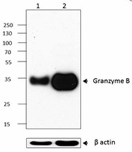
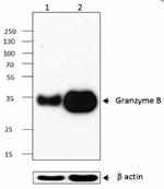
Western blot analysis of PBMC (lane 1), PBMC activated with 5 µg/ml CD3 antibody coated on the plate and 2 µg/ml CD28 antibody for two days (lane 2) using anti-Granzyme B antibody (clone M3304B06). Anti-β actin antibody (poly6221) was used as a loading control. -

IHC staining of purified anti-Granzyme B antibody (clone M3304B06) on formalin-fixed paraffin-embedded human tonsil tissue. Following antigen retrieval using Tris-EDTA buffer (10mM Tris, 1mM EDTA, pH 9.0), the tissue was incubated with 5.0 µg/mL of the primary antibody overnight at 4&Deg;C, followed by incubation with Alexa Fluor® 647 goat anti-mouse IgG (magenta) (Cat. No. 405322) for one hour at room temperature. Nuclei were counterstained with DAPI (blue) (Cat. No. 422801), and the slides were mounted with ProLong™ Gold Antifade Mountant. Scale bar: 50 µm. B: Crop image of A. Scale bar: 10 µm -

SeqIF™ (sequential immunofluorescence) staining on COMET™ of Purified anti-Granzyme B (clone M3304B06, yellow) on formalin-fixed paraffin-embedded human tonsil tissue at 5 µg/mL. Alexa Fluor™ Plus 647 Goat anti-Mouse IgG antibody (Lunaphore, Cat. No. DR647MS) was used as a secondary antibody. Nuclei were counterstained with DAPI (blue). Tissue underwent an all-in-one dewaxing and antigen retrieval preprocessing.
| Cat # | Size | Price | Quantity Check Availability | ||
|---|---|---|---|---|---|
| 674602 | 100 µg | $300.00 | |||
Granzyme B is a serine protease that is mainly produced by cytotoxic T cells (CTL) and NK cells. It plays important roles in cytotoxic lymphocyte-mediated apoptosis, chronic inflammation, and impaired wound healing. Granzyme B is packaged in cytoplasmic granules that are exocytosed towards a bound target cell, and subsequently activates multiple protein substrates to induce apoptosis. Perforin facilitates the transfer of Granzyme B into the target cell. Most circulating CD56+ CD8- NK cells and approximately half of circulating CD8+ T cells coexpress both Granzyme A and B. The activation of CD8+ and CD4+ T lymphocytes induces substantial expression of Granzyme B, but not Granzyme A. Recently, Granzyme B has been found to be produced by other cell types such as CD34+ hematopoietic progenitor cells, keratinocytes, basophils, mast cells, plasmacytoid dendritic cells, B cells, and smooth muscle cells. Elevated levels of Granzyme B are also found in some kinds of autoimmune diseases, type 1 diabetes, and cardiovascular diseases.
Product Details
- Verified Reactivity
- Human
- Antibody Type
- Monoclonal
- Host Species
- Mouse
- Immunogen
- Recombinant human Granzyme B produced in the 293E cell line.
- Formulation
- Phosphate-buffered solution, pH 7.2, containing 0.09% sodium azide.
- Preparation
- The antibody was purified by affinity chromatography.
- Concentration
- 0.5 mg/mL
- Storage & Handling
- The antibody solution should be stored undiluted between 2°C and 8°C.
- Application
-
WB - Quality tested
IHC-P - Verified
SB - Community verified - Recommended Usage
-
Each lot of this antibody is quality control tested by Western blotting. For Western blotting, the suggested use of this reagent is 0.5 - 2.0 µg per mL. For immunohistochemistry on formalin-fixed paraffin-embedded tissue sections, a concentration range of 1.0 - 10 µg/mL is suggested. It is recommended that the reagent be titrated for optimal performance for each application.
- Additional Product Notes
-
For the use of this antibody in spatial biology applications, we have partnered with Lunaphore Technologies for demonstration of our antibodies on the COMET™. The COMET™ platform is an automated, end-to-end spatial biology solution developed for rapid and flexible multiplex tissue profiling. More information on the COMET™ and a complete list of our antibodies that have been demonstrated on the COMET™ can be found here.
- Product Citations
-
- RRID
-
AB_2565266 (BioLegend Cat. No. 674602)
Antigen Details
- Structure
- 247 amino acids with a molecular weight of approximately 28 kD
- Distribution
-
Cytotoxic T cells, NK cells, and neutrophils.
- Function
- Granzyme B is able to induce target cell apoptosis by activating caspase independent pathways. Granzyme B is induced in CD8+ T lymphocytes with ConA / IL-2 and CD4+ T lymphocytes with anti-CD3/CD28 or anti-CD3/CD46.
- Interaction
- Targets of CTL and NK cells.
- Cell Type
- B cells, Dendritic cells, Neutrophils, NK cells, T cells
- Biology Area
- Apoptosis/Tumor Suppressors/Cell Death, Cell Biology, Cell Cycle/DNA Replication, Immunology, Innate Immunity, Neuroscience, Signal Transduction
- Molecular Family
- Enzymes and Regulators, Proteases
- Antigen References
-
1. Edwards KM, et al. 1999. J. Biol. Chem. 274:30468.
2. Grossman WJ, et al. 2004. Blood 104:2840.
3. Heusel JW, et al. 1994. Cell 76:977.
4. Schmid J and Weissmann C. 1987. J. Immunol. 139:250.
5. Trapani JA, et al. 1988. Proc. Natl. Acad. Sci. USA 85:6924.
6. Hiebert PR and Granville DJ. 2012. Trends Mol. Med. 18:732.
7. Saito Y, et al. 2011. J. Cardiol. 57:141.
8. Ewen CL, et al. 2012. Cell Death Differ. 1:28. - Gene ID
- 3002 View all products for this Gene ID
- UniProt
- View information about Granzyme B on UniProt.org
Other Formats
View All Granzyme B Reagents Request Custom Conjugation| Description | Clone | Applications |
|---|---|---|
| Purified anti-Granzyme B | M3304B06 | WB,IHC-P,SB |
| TotalSeq™-Bn1337 anti-Granzyme B | M3304B06 | SB |
Compare Data Across All Formats
This data display is provided for general comparisons between formats.
Your actual data may vary due to variations in samples, target cells, instruments and their settings, staining conditions, and other factors.
If you need assistance with selecting the best format contact our expert technical support team.
-
Purified anti-Granzyme B

Western blot analysis of PBMC (lane 1), PBMC activated with ... 
IHC staining of purified anti-Granzyme B antibody (clone M33... 
SeqIF™ (sequential immunofluorescence) staining on COMET™ of... -
TotalSeq™-Bn1337 anti-Granzyme B
