- Clone
- C398.4A (See other available formats)
- Regulatory Status
- RUO
- Other Names
- Inducible COStimulatory molecule, H4
- Isotype
- Armenian Hamster IgG
-

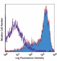
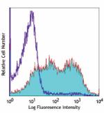
PHA-stimulated human peripheral blood lymphocytes (3 days) stained with purified C398.4A, followed by biotinylated anti-Armenian hamster IgG and Sav-PE -

SeqIF™ (sequential immunofluorescence) staining on COMET™ of Purified anti-CD278 (ICOS) (clone C398.4A, yellow) on formalin-fixed paraffin-embedded human tonsil tissue at 5 µg/mL. Alexa Fluor® 647 Goat anti-Armenian hamster IgG antibody was used as secondary antibody. Nuclei were counterstained with DAPI (blue). Tissue underwent an all-in-one dewaxing and antigen retrieval preprocessing.
| Cat # | Size | Price | Quantity Check Availability | ||
|---|---|---|---|---|---|
| 313502 | 500 µg | $270.00 | |||
ICOS, also known as inducible costimulatory molecule and H4, is a 47-57 kD protein. This protein is homologous to the CD28/CTLA-4 proteins. ICOS is expressed on activated T cells and a subset of thymocytes. It is able to costimulate T cells proliferation. In addition, ICOS is involved in humoral immune responses (B cell germinal center formation). The ICOS ligand is B7h/B7RP-1 or B7-H2. ICOS stimulation has been shown to potentiate TCR-mediated IL-4 and IL-10 production and has been proposed to play a role in Th2 cell development.
Product Details
- Verified Reactivity
- Human, Mouse, Rat
- Reported Reactivity
- African Green, Baboon, Cynomolgus, Rhesus, Pig
- Antibody Type
- Monoclonal
- Host Species
- Armenian Hamster
- Immunogen
- Mouse T cell clone D10.G4.1
- Formulation
- Phosphate-buffered solution, pH 7.2, containing 0.09% sodium azide.
- Preparation
- The antibody was purified by affinity chromatography.
- Concentration
- 0.5 mg/ml
- Storage & Handling
- The antibody solution should be stored undiluted between 2°C and 8°C.
- Application
-
FC - Quality tested
IP, Costim - Reported in the literature, not verified in house
SB - Community verified - Recommended Usage
-
Each lot of this antibody is quality control tested by immunofluorescent staining with flow cytometric analysis. For flow cytometric staining, the suggested use of this reagent is ≤ 1.0 µg per 106 cells in 100 µl volume. It is recommended that reagents be titrated for optimal performance in the particular application.
- Application Notes
-
- The C398.4A antibody is useful for flow cytometric analysis and is able to costimulate T cell activation and proliferation. Additional reported applications (for the relevant formats) include: immunoprecipitation1, in vitro costimulation of T cell activation1,3,4, and spatial biology (IBEX)5,6. The LEAF™ purified antibody (Endotoxin < 0.1 EU/µg, Azide-Free, 0.2 µm filtered) is recommended for functional assays (Cat. No. 313512).
- Additional Product Notes
-
For the use of this antibody in spatial biology applications, we have partnered with Lunaphore Technologies for demonstration of our antibodies on the COMET™. The COMET™ platform is an automated, end-to-end spatial biology solution developed for rapid and flexible multiplex tissue profiling. More information on the COMET™ and a complete list of our antibodies that have been demonstrated on the COMET™ can be found here.
-
Application References
(PubMed link indicates BioLegend citation) -
- Redoglia V, et al. 1996. Eur. J. Immunol. 26:2781. (FC IP Costim)
- Yagi J, et al. 2003. J. Immunol. 171:783. (FC)
- Arimura Y, et al. 2002. Int. Immunol. 14:555. (Costim)
- Arimura Y, et al. 2004. J. Biol. Chem. 279:11408. (Costim)
- Radtke AJ, et al. 2020. Proc Natl Acad Sci USA. 117:33455-33465. (SB) PubMed
- Radtke AJ, et al. 2022. Nat Protoc. 17:378-401. (SB) PubMed
- Product Citations
-
- RRID
-
AB_416326 (BioLegend Cat. No. 313502)
Antigen Details
- Structure
- CD28/CTLA-4, 47-57 kD
- Distribution
-
Activated T cells, subset of thymocytes
- Function
- Costimulates T cell activation, proliferation, humoral immune response
- Ligand/Receptor
- B7h/B7RP-1/GL-50
- Cell Type
- T cells, Thymocytes, Tregs
- Biology Area
- Costimulatory Molecules, Immunology
- Molecular Family
- CD Molecules
- Antigen References
-
1. Redoglia V, et al. 1996. Eur. J. Immunol. 26:2781.
2. Hutloff A, et al. 1999. Nature 397:263.
3. Buonfiglio D, et al. 2000. Eur. J. Immunol. 30:3463.
4. Coyle AJ, et al. 2000. Immunity 13:95. - Gene ID
- 100048841 View all products for this Gene ID 29851 View all products for this Gene ID 64545 View all products for this Gene ID
- UniProt
- View information about CD278 on UniProt.org
Other Formats
View All CD278 Reagents Request Custom ConjugationCompare Data Across All Formats
This data display is provided for general comparisons between formats.
Your actual data may vary due to variations in samples, target cells, instruments and their settings, staining conditions, and other factors.
If you need assistance with selecting the best format contact our expert technical support team.
-
Purified anti-human/mouse/rat CD278 (ICOS)
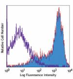
PHA-stimulated human peripheral blood lymphocytes (3 days) s... 
SeqIF™ (sequential immunofluorescence) staining on COMET™ of... -
Biotin anti-human/mouse/rat CD278 (ICOS)
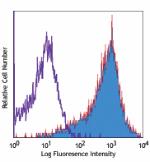
PHA-stimulated human peripheral blood lymphocytes (3 days) s... -
FITC anti-human/mouse/rat CD278 (ICOS)
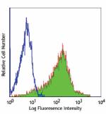
PHA-stimulated human peripheral blood lymphocytes (3 days) s... -
PE anti-human/mouse/rat CD278 (ICOS)
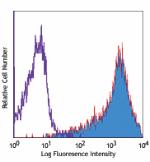
PHA-stimulated human peripheral blood lymphocytes (3 days) s... -
APC anti-human/mouse/rat CD278 (ICOS)
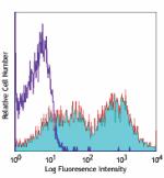
PHA-stimulated human peripheral blood lymphocytes (3 days) s... -
Alexa Fluor® 488 anti-human/mouse/rat CD278 (ICOS)
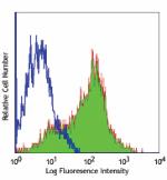
PHA-stimulated human peripheral blood lymphocytes (3 days) s... 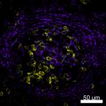
Confocal image of human lymph node sample acquired using the... -
Alexa Fluor® 647 anti-human/mouse/rat CD278 (ICOS)
PHA-stimulated human peripheral blood lymphocytes (3 days) s... 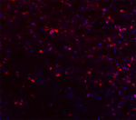
Frozen mouse lymph node section detected with anti-ICOS Alex... -
PerCP/Cyanine5.5 anti-human/mouse/rat CD278 (ICOS)
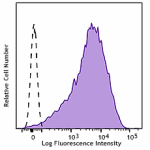
PHA-stimulated (three days) human peripheral blood lymphocyt... -
PE/Cyanine7 anti-human/mouse/rat CD278 (ICOS)
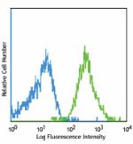
PHA-stimulated human peripheral blood lymphocytes (3 days) s... -
Pacific Blue™ anti-human/mouse/rat CD278 (ICOS)
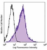
PHA-activated (3 days) human peripheral blood lymphocytes we... -
Brilliant Violet 421™ anti-human/mouse/rat CD278 (ICOS)
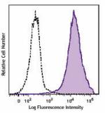
PHA-activated (3 days) human peripheral blood lymphocytes we... -
Brilliant Violet 510™ anti-human/mouse/rat CD278 (ICOS)
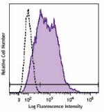
PHA-activated (3 days) human peripheral blood lymphocytes we... -
APC/Cyanine7 anti-human/mouse/rat CD278 (ICOS)
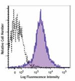
PHA-activated (three days) human peripheral blood lymphocyte... -
PE/Dazzle™ 594 anti-human/mouse/rat CD278 (ICOS)
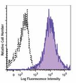
PHA-activated (three days) human peripheral blood lymphocyte... -
Alexa Fluor® 700 anti-human/mouse/rat CD278 (ICOS)
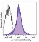
PHA-activated (three days) human peripheral blood lymphocyte... -
APC/Fire™ 750 anti-human/mouse/rat CD278 (ICOS)
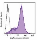
PHA-activated (three days) human peripheral blood lymphocyte... -
Brilliant Violet 785™ anti-human/mouse/rat CD278 (ICOS)
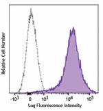
PHA-activated (three days) human peripheral blood lymphocyte... -
Brilliant Violet 605™ anti-human/mouse/rat CD278 (ICOS)
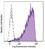
PHA-stimulated human peripheral blood lymphocytes (3 days) s... -
Ultra-LEAF™ Purified anti-human/mouse/rat CD278 (ICOS)
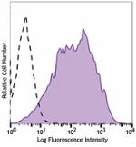
PHA-stimulated human peripheral blood lymphocytes (3 days) w... -
GoInVivo™ Purified anti-human/mouse/rat CD278 (ICOS)
-
Brilliant Violet 711™ anti-human/mouse/rat CD278 (ICOS)
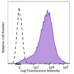
PHA-stimulated human peripheral blood lymphocytes (3 days) s... -
Brilliant Violet 650™ anti-human/mouse/rat CD278 (ICOS)
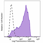
PHA-stimulated human peripheral blood lymphocytes (3 days) s... -
TotalSeq™-B0171 anti-human/mouse/rat CD278 (ICOS)
-
TotalSeq™-C0171 anti-human/mouse/rat CD278 (ICOS)
-
TotalSeq™-A0171 anti-human/mouse/rat CD278 (ICOS)
-
Brilliant Violet 750™ anti-human/mouse/rat CD278 (ICOS)
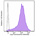
PHA-stimulated (3 days) human peripheral blood lymphocytes w... -
TotalSeq™-D0171 anti-human/mouse/rat CD278 (ICOS) Antibody
-
PE/Cyanine5 anti-human/mouse/rat CD278 (ICOS)
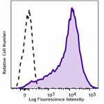
PHA stimulated human peripheral blood lymphocytes (3 days) w... -
Spark Red™ 718 anti-human/mouse/rat CD278 (ICOS)
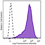
PHA-stimulated (three days) human peripheral blood lymphocyt...
