- Clone
- W19210B (See other available formats)
- Regulatory Status
- RUO
- Other Names
- Hermes, Pgp-1, H-CAM, HUTCH-1, ECMR III, gp85, Ly-24
- Isotype
- Rat IgG2a, κ

-

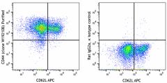
Mouse splenocytes were stained with anti-mouse CD62L APC and purified anti-mouse CD44 (clone W19210B) (left) or rat IgG2a, κ isotype control (right) followed by PE anti-rat IgG. -

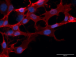
ICC staining of purified anti-mouse CD44 (clone W19210B) on NIH-3T3 cells. The cells were fixed with 4% PFA, and permeabilized with 100% ice-cold methanol. Cells were then blocked with 5% fetal bovine serum in PBS for 30 minutes, and then incubated with 5 µg/mL of the primary antibody overnight at 4°C, followed by incubation with 2.5 µg/mL of Alexa Fluor® 594 goat anti-rat IgG (red) (Cat. No. 405422) for one hour at room temperature. Nuclei were counterstained with DAPI (blue), and the slides were mounted with ProLong™ Gold Antifade Mountant. The image was captured with a 40X objective. Scale bar: 50 µm -

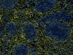
IHC staining using purified anti-mouse CD44 (clone W19210B) on formalin-fixed frozen mouse spleen tissue. The tissue was incubated with 5 μg/mL of the primary antibody overnight at 4°C, followed by incubation with 2.5 μg/mL of Alexa Fluor® 647 goat anti-rat IgG (yellow) (Cat. No. 405416) for one hour at room temperature. Nuclei were counterstained with DAPI (blue), and the slide was mounted with ProLong™ Gold Antifade Mountant. The image was captured with a 10x objective.
| Cat # | Size | Price | Quantity Check Availability | ||
|---|---|---|---|---|---|
| 949601 | 25 µg | $118.00 | |||
| 949602 | 100 µg | $293.00 | |||
CD44 is a 80-95 kD glycoprotein also known as Hermes, Pgp1, H-CAM, or HUTCH. It is expressed on all leukocytes, endothelial cells, hepatocytes, and mesenchymal cells. As B and T cells become activated or progress to the memory stage, CD44 expression increases from low or mid levels to high levels. Thus, CD44 has been reported to be a valuable marker for memory cell subsets. High CD44 expression on Treg cells has been associated with potent suppressive function via high production of IL-10. CD44 is an adhesion molecule involved in leukocyte attachment to and rolling on endothelial cells, homing to peripheral lymphoid organs and to the sites of inflammation, and leukocyte aggregation.
Product Details
- Verified Reactivity
- Mouse
- Antibody Type
- Monoclonal
- Host Species
- Rat
- Immunogen
- Partial recombinant mouse CD44 protein
- Formulation
- Phosphate-buffered solution, pH 7.2, containing 0.09% sodium azide
- Preparation
- The antibody was purified by affinity chromatography.
- Concentration
- 0.5 mg/mL
- Storage & Handling
- The antibody solution should be stored undiluted between 2°C and 8°C.
- Application
-
FC - Quality tested
ICC, IHC-F - Verified - Recommended Usage
-
Each lot of this antibody is quality control tested by immunofluorescent staining with flow cytometric analysis. For flow cytometric staining, the suggested use of this reagent is ≤ 0.25 µg per million cells in 100 µL volume. For immunocytochemistry, a concentration range of 2.5 - 5.0 μg/mL is recommended. For immunohistochemical staining on frozen tissue sections, a concentration range of 1.125 - 5.0 µg/mL is suggested It is recommended that the reagent be titrated for optimal performance for each application.
- Application Notes
-
Not recommended for western blotting. Not reactive for human CD44 (tested in WB and IHC-P).
- RRID
-
AB_2922650 (BioLegend Cat. No. 949601)
AB_2922650 (BioLegend Cat. No. 949602)
Antigen Details
- Structure
- Variable splicing of CD44 gene generates many CD44 isoforms, 80-95 kD
- Distribution
-
All leukocytes, epithelial cells, endothelial cells, hepatocytes, mesenchymal cells
- Function
- Leukocyte attachment and rolling on endothelial cells, stromal cells and ECM
- Ligand/Receptor
- Hyaluronan, MIP-1β, fibronectin, collagen
- Cell Type
- B cells, Endothelial cells, Epithelial cells, Leukocytes, Mesenchymal cells
- Biology Area
- Immunology
- Molecular Family
- Adhesion Molecules, CD Molecules
- Antigen References
-
- Barclay AN, et al. 1997. The Leukocyte Antigen FactsBook Academic Press.
- Haynes BF, et al. 1991. Cancer Cells. 3:347-50.
- Goldstein LA, et al. 1989. Cell. 56:1063-72.
- Mikecz K, et al. 1995. Nat Med. 1:558-63.
- Hegde V, et al. 2008. J Leukoc Biol. 84:134-42.
- Liu T, et al. 2009. Biol Direct. 4:40.
- Gene ID
- 960 View all products for this Gene ID
- UniProt
- View information about CD44 on UniProt.org
Other Formats
View All CD44 Reagents Request Custom Conjugation| Description | Clone | Applications |
|---|---|---|
| Purified anti-mouse CD44 | W19210B | FC,ICC,IHC-F |
Compare Data Across All Formats
This data display is provided for general comparisons between formats.
Your actual data may vary due to variations in samples, target cells, instruments and their settings, staining conditions, and other factors.
If you need assistance with selecting the best format contact our expert technical support team.


