- Clone
- 12H6 (See other available formats)
- Regulatory Status
- RUO
- Other Names
- Cadherin 3, CDH3, Placental Cadherin
- Isotype
- Mouse IgG1, κ

-

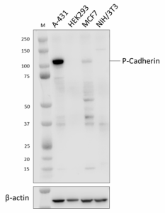
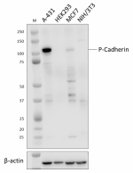
Whole cell extracts (15 µg total protein) from A-431, HEK293, MCF7, and NIH/3T3 cells were resolved by 4-12% Bis-Tris gel electrophoresis, transferred to a PVDF membrane, and probed with 1.0 μg/mL of purified anti-P-Cadherin antibody (clone 12H6) overnight at 4 °C. Proteins were visualized by chemiluminescence detection using HRP goat anti-mouse IgG antibody (Cat. No. 405306) at a 1:3000 dilution. Direct-Blot™ HRP anti-β-actin antibody (Cat. No. 643807) was used as a loading control at a 1:10,000 dilution (lower). Lane M: Molecular weight marker. -

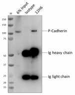
Whole cell extracts (250 µg total protein) prepared from A31 cells were immunoprecipitated overnight with 2.5 µg of purified mouse IgG1, κ isotype ctrl antibody (Cat. No. 400102) or purified anti-P-Cadherin antibody (clone 12H6). The resulting IP fractions and whole cell extract input (6%) were resolved by 4-12% Bis-Tris gel electrophoresis, transferred to a PVDF membrane, and probed with a mouse control antibody. Lane M: Molecular weight marker.
| Cat # | Size | Price | Quantity Check Availability | ||
|---|---|---|---|---|---|
| 948401 | 25 µg | $118.00 | |||
| 948402 | 100 µg | $293.00 | |||
P-cadherin, or placental-cadherin, belongs to one of the three subclasses in the cadherin family. Cadherins are Ca2+ dependent adherent proteins involved in intercellular cell adhesions. P-cadherin contains a cytoplasmic carboxy-terminal domain, a transmembrane domain, and a extracellular N-terminal domain. P-cadherin was first identified in mouse placental tissue. However, in humans, the distribution of P-cadherin is restricted to the basal or lower layers of the stratified epithelia in the epithelial layer, and is not found in human placental tissue. In humans, the loss of functional P-cadherin can induce characteristic genetic syndromes such as hypotrichosis with juvenile muscular dystrophy (HJMD) and ectodermal dysplasia, ectrodactyly, and muscular dystrophy (EEM syndrome). In breast cancer, an overexpression of P-cadherin is often found in high histological grade tumors. In an in vitro study, the overexpression of P-cadherin in breast cancer cells in the presence of wild type E-cadherin, promoted cell invasiveness, motility, and migration.
Product Details
- Verified Reactivity
- Human
- Antibody Type
- Monoclonal
- Host Species
- Mouse
- Immunogen
- Recombinant full-length Human P-cadherin peptide.
- Formulation
- Phosphate-buffered solution, pH 7.2, containing 0.09% sodium azide
- Preparation
- The antibody was purified by affinity chromatography.
- Concentration
- 0.5 mg/mL
- Storage & Handling
- The antibody solution should be stored undiluted between 2°C and 8°C.
- Application
-
WB - Quality tested
IP - Verified - Recommended Usage
-
Each lot of this antibody is quality control tested by western blotting. For western blotting, the suggested use of this reagent is 0.5 - 1.0 µg/mL. For immunoprecipitation, the suggested use of this reagent is 2.5 µg/test. It is recommended that the reagent be titrated for optimal performance for each application.
- Application Notes
-
This clone was tested for ICC using 4% PFA-fixed A-431 cells permeabilized with either methanol or Triton X-100. Neither method was compatible with P-Cadherin staining. This clone was also tested using methanol fixation on A-431 and HaCaT cells. Neither cell line was compatible with P-Cadherin staining.
- RRID
-
AB_2894538 (BioLegend Cat. No. 948401)
AB_2894538 (BioLegend Cat. No. 948402)
Antigen Details
- Structure
- P-Cadherin is a 829 amino acid protein with a predicted molecular weight of 91 kD.
- Distribution
-
Adhesion protein/Plasma membrane
- Function
- Intercellular cell adhesion
- Biology Area
- Cell Adhesion, Cell Biology, Cell Motility/Cytoskeleton/Structure
- Antigen References
-
- Ribeiro A. et al. 2010. Oncogene. 29:392-402.
- Shimoyama Y. and Hirohashi S. 1991. Cancer Research. 51:2185-2192.
- Sun L. et al. 2011. American Journal of Pathology. 179:380-390.
- Vieira A. & Paredes J. 2015. Molecular Cancer. 14:178.
- Gene ID
- 1001 View all products for this Gene ID
- UniProt
- View information about P-Cadherin on UniProt.org
Other Formats
View All P-Cadherin Reagents Request Custom Conjugation| Description | Clone | Applications |
|---|---|---|
| Purified anti-P-Cadherin | 12H6 | WB,IP |
Compare Data Across All Formats
This data display is provided for general comparisons between formats.
Your actual data may vary due to variations in samples, target cells, instruments and their settings, staining conditions, and other factors.
If you need assistance with selecting the best format contact our expert technical support team.


