- Clone
- A20103O (See other available formats)
- Regulatory Status
- RUO
- Other Names
- S1, Spike Protein
- Isotype
- Rat IgG2a, κ

-

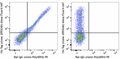
His-tagged SARS-CoV-2 S protein S1 transfected CHO cells were stained with anti-S1 (clone A20103O) purified (left) or rat IgG2a, κ isotype control (clone RTK2758) purified (right) followed by anti-rat IgG PE (clone Poly4054) and Alexa Fluor® 647 anti-His Tag (clone J095G46). -

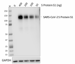
Whole cell extracts (15 µg total protein) from HeLa cells mixed with the indicated amount of His-tagged recombinant SARS-CoV-2 S Protein S1 (Cat. No. 792906) were resolved by 4-12% Bis-Tris gel electrophoresis, transferred to a PVDF membrane, and probed with 1.0 µg/mL (1:500 dilution) purified anti-SARS-CoV-2 S Protein S1 antibody (clone A20103O) overnight at 4°C. Proteins were visualized by chemiluminescence detection using HRP goat anti-human IgG antibody at a 1:3000 dilution. Direct-Blot™ HRP anti-GAPDH antibody (Cat. No. 607904) was used as a loading control at a 1:25000 dilution (lower). Lane M: Molecular weight marker. -

200 ng of recombinant SARS-CoV-2 S protein S1 + S2 (Cat. No. 793704) was coated onto a Costar™ 96-well high binding assay plate and incubated with a dilution series of purified anti-SARS-CoV-2 S protein S1 antibody (clone A20103O). Bound antibodies were detected with HRP goat anti-mouse IgG antibody (Cat. No. 405306) followed by TMB substrate solution. Absorbance was measured at 450 nm.
| Cat # | Size | Price | Quantity Check Availability | ||
|---|---|---|---|---|---|
| 945101 | 25 µg | $112.00 | |||
| 945102 | 100 µg | $335.00 | |||
SARS-CoV-2 is a respiratory virus which causes coronavirus disease 2019 (COVID-19). The coronavirus spike (S) glycoprotein is a class I viral fusion protein on the outer envelope of the virion that plays a critical role in viral infection by recognizing host cell receptors and mediating fusion of the viral and cellular membranes. The S glycoprotein is synthesized as a precursor protein consisting of ~1,300 amino acids that is then cleaved into an amino (N)-terminal S1 subunit (~700 amino acids) and a carboxyl (C)-terminal S2 subunit (~600 amino acids). Three S1/S2 heterodimers assemble to form a trimer spike protruding from the viral envelope. The S1 subunit contains a receptor-binding domain (RBD) that can specifically bind to angiotensin-converting enzyme 2 (ACE2), the receptor on target cells. Triggered by receptor binding, proteolytic processing and/or acidic pH in the cellular compartments, the class I viral fusion protein undergoes a transition from a metastable pre-fusion state to a stable post-fusion state during infection, in which the receptor-binding subunit is cleaved, and the fusion subunit undergoes large-scale conformational rearrangements to expose the hydrophobic fusion peptide, induce the formation of a six-helix bundle, and bring the viral and cellular membranes close for fusion. The trimeric SARS coronavirus (SARS-CoV-2) S glycoprotein consisting of three S1-S2 heterodimers binds the cellular receptor angiotensin-converting enzyme 2 (ACE2) and mediates fusion of the viral and cellular membranes through a pre- to post-fusion conformation transition.
Product Details
- Verified Reactivity
- SARS-CoV-2
- Antibody Type
- Monoclonal
- Host Species
- Rat
- Immunogen
- Partial recombinant SARS-CoV-2 S protein corresponding to S1 subunit
- Formulation
- Phosphate-buffered solution, pH 7.2, containing 0.09% sodium azide
- Preparation
- The antibody was purified by affinity chromatography.
- Concentration
- 0.5 mg/mL
- Storage & Handling
- The antibody solution should be stored undiluted between 2°C and 8°C.
- Application
-
WB - Quality tested
Direct ELISA, FC - Verified - Recommended Usage
-
Each lot of this antibody is quality control tested by western blotting. For western blotting, the suggested use of this reagent is 1.0 µg/mL. For Direct ELISA, a concentration of 114.9 ng/mL is recommended. For flow cytometric staining, the suggested use of this reagent is ≤ 0.5 µg per million cells in 100 µL volume. It is recommended that the reagent be titrated for optimal performance for each application.
- Application Notes
-
BioLegend offers multiple clones that recognize SARS-CoV-2 S protein S1. Clone A20103H displayed the strongest performance for western blot of all clones validated for the application.
- Product Citations
-
- RRID
-
AB_2890875 (BioLegend Cat. No. 945101)
AB_2890875 (BioLegend Cat. No. 945102)
Antigen Details
- Structure
- Spike glycoprotein is a homotrimer. Each monomer consists of 1,273 amino acids with a theoretical molecular weight of 141 kD, and consists of S1 and S2 subunits.
- Distribution
-
Viral envelope protein, host cell membrane, host cell endoplasmic reticulum-Golgi intermediate compartment membrane
- Function
- Mediates fusion between virus and host cell membranes
- Interaction
- ACE2
- Ligand/Receptor
- ACE2
- Biology Area
- COVID-19
- Antigen References
-
1. Walls AC, et al. 2020. Cell. 181(2):281-292.
2. Yan R, et al. 2020. Science. 367 (6485):1444-1448.
3. Wrapp D, et al. 2020. Science. 367 (6483):1260-1263.
4. Shang J, et al. 2020. PNAS. 117(21):11727-11734 - Gene ID
- 43740568 View all products for this Gene ID
- UniProt
- View information about Viral Protein on UniProt.org
Other Formats
View All 945101 Reagents Request Custom Conjugation| Description | Clone | Applications |
|---|---|---|
| Purified anti-SARS-CoV-2 S Protein S1 Antibody | A20103O | WB,FC,ELISA |
Compare Data Across All Formats
This data display is provided for general comparisons between formats.
Your actual data may vary due to variations in samples, target cells, instruments and their settings, staining conditions, and other factors.
If you need assistance with selecting the best format contact our expert technical support team.
-
Purified anti-SARS-CoV-2 S Protein S1 Antibody

His-tagged SARS-CoV-2 S protein S1 transfected CHO cells wer... 
Whole cell extracts (15 µg total protein) from HeLa cells mi... 
200 ng of recombinant SARS-CoV-2 S protein S1 + S2 (Cat. No....
