- Clone
- H76 (See other available formats)
- Regulatory Status
- RUO
- Other Names
- Lymphocyte cytoplasmic protein 2 (LCP2), SH2 domain containing leukocyte 76kD protein
- Isotype
- Mouse IgG2a, κ

-

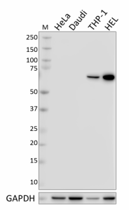
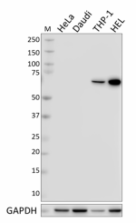
Total cell lysates (15 µg total protein) from HeLa (negative control), Daudi (negative control), THP-1, and HEL cells were resolved by 4-12% Bis-Tris gel electrophoresis, transferred to a nitrocellulose membrane, and probed with 0.5 µg/mL (1:1000 dilution) of purified anti-SLP-76 antibody, clone H76, overnight at 4°C. Proteins were visualized by chemiluminescence detection using HRP goat anti-mouse IgG (Cat. No. 405301) at a 1:3000 dilution. Direct-Blot™ HRP anti-GAPDH Antibody (Cat. No. 664804) was used as a loading control at a 1:25000 dilution (lower). Lane M: Molecular Weight marker. Predicted expression data was obtained from Human Protein Atlas. -

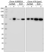
Whole cell extracts (15 µg protein) from serum starved Jurkat and EL4 cells untreated (-) or treated (+) with 5 mM H2O2 were resolved on a 4-12% Bis-Tris gel, transferred to nitrocellulose and probed with 0.1 µg/mL (1:5000 dilution) of purified anti-SLP76 Phospho (Ser376), clone A18001A (Cat. No. 667102), for 2 hours at room temperature. Proteins were visualized by chemiluminescence detection using HRP goat anti-mouse IgG antibody (Cat. No. 405301) at a 1:3000 dilution. Equal SLP76 loading was confirmed by probing membranes with anti-pan SLP76 antibody, clone H76 (Cat. No. 625002) at a 1:1000 dilution. Lane M: Molecular Weight marker.
| Cat # | Size | Price | Quantity Check Availability | ||
|---|---|---|---|---|---|
| 625002 | 200 µL | $293.00 | |||
SLP-76 is a human Src homology domain-containing leukocyte protein. This cytoplasmic adaptor phosphoprotein contains both SH2 and SAM domains and is involved in B and T cell receptor signaling. The amino terminus of this protein contains three 17 amino acid repeats with conserved tyrosine and acidic residues (DYE(S/P)P) as well as a proline rich region and is known to associate with many proteins involved in T and B cell signaling including Grb2, LAT, Vav1, SLAP-130, SHP-1, and phospholipase C gamma 2. SLP-76 can be phosphorylated on multiple tyrosine residues by the upstream kinases ZAP-70 and Lck. SLP-76 phosphorylation plays an important role in T cell-mediated IL-2 production by allowing phosphorylated Vav to bind; this complex (SLP-76/Vav) stimulates NF-AT and IL-2 gene activation after TCR engagement. Overexpression of SLP-76 has been shown to result in enhanced IL-2 transcription after TCR signaling. The H76 monoclonal antibody recognizes human SLP-76 and has been shown to be useful for Western blotting.
Product Details
- Verified Reactivity
- Human, Mouse
- Antibody Type
- Monoclonal
- Host Species
- Mouse
- Immunogen
- amino acids 216-434 of human SLP-76
- Formulation
-
This antibody is provided in phosphate-buffered solution, containing 0.09% sodium azide.
Previous lots of this product may have been formulated with 0.1% or 0.05% NaN3 instead of 0.09% NaN3. For further information please contact BioLegend Technical Support or Customer Service. - Preparation
- The antibody was purified by affinity chromatography.
- Concentration
- Lot-specific (to obtain lot-specific concentration, please enter the lot number in our Concentration and Expiration Lookup or Certificate of Analysis online tools.)
- Storage & Handling
- Upon receipt, aliquot and store frozen at -20°C. Avoid freeze-thaw cycles.
- Application
-
WB - Quality tested
IP - Reported in the literature, not verified in house - Recommended Usage
-
Each lot of this antibody is quality control tested by Western blotting. Western blotting, suggested working dilution(s): Use 1:1000 dilution buffer for each mini-gel. It is recommended that the reagent be titrated for optimal performance for each application.
- Additional Product Notes
-
The 200 µL size can be used for approximately 40 Western blots.
-
Application References
(PubMed link indicates BioLegend citation) -
- Deford-Watts LM, et al. 2011. J. Immunol. 186:6839. PubMed.
- Product Citations
-
- RRID
-
SCR_001134 (BioLegend Cat. No. 625002)
Antigen Details
- Structure
- Phosphoprotein 76 kD containing SH2 domain in the C-terminus
- Distribution
-
Expressed in leukocytes (monocytes, T and B cells), thymus, spleen, placenta, and lung.
- Function
- Adaptor molecule involved in B cell receptor and T cell receptor signaling
- Interaction
- Grb2, LAT, Vav1, SLAP-130, SHP-1, phospholipase C gamma 2
- Modification
- Phosphorylation (Y113, Y128, Y145, Y423, Y426)
- Cell Type
- B cells, Monocytes, T cells
- Biology Area
- Cell Biology, Immunology, Signal Transduction
- Molecular Family
- Protein Kinases/Phosphatase
- Antigen References
-
1. Jackman KJ, et al. 1995. J. Biol. Chem. 270:7029.
2. Wardenberg JB, et al. 1996. J. Biol. Chem. 271:19641.
3. Tuosto L, et al. 1996. J. Exp. Med. 184:1161.
4. Motto DG, et al. 1996. J. Exp. Med. 183:1937. - Gene ID
- 3937 View all products for this Gene ID
- UniProt
- View information about SLP-76 on UniProt.org
Other Formats
View All SLP-76 Reagents Request Custom Conjugation| Description | Clone | Applications |
|---|---|---|
| Purified anti-SLP-76 | H76 | WB,IP |
Compare Data Across All Formats
This data display is provided for general comparisons between formats.
Your actual data may vary due to variations in samples, target cells, instruments and their settings, staining conditions, and other factors.
If you need assistance with selecting the best format contact our expert technical support team.


