- Clone
- QA20B02 (See other available formats)
- Regulatory Status
- RUO
- Other Names
- Spleen Associated Tyrosine Kinase, Spleen Tyrosine Kinase, Tyrosine-Protein Kinase Syk, IMD82
- Isotype
- Mouse IgG2b, κ

-

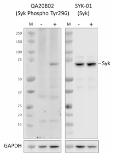
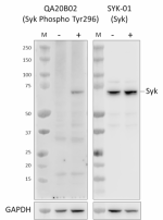
Whole cell extracts (15 µg protein) from serum-starved Ramos cells untreated (-) or treated (+) with 5 mM H2O2 for 3 minutes were resolved on a 4-12% Bis-Tris gel, transferred to a PVDF membrane, and probed with 1.0 µg/mL (1:500 dilution) of purified anti-Syk Phospho (Tyr296) recombinant (clone QA20B02) for 2 hours at room temperature. Proteins were visualized by chemiluminescence detection using HRP goat anti-mouse IgG (Cat. No. 405306) at a 1:3000 dilution. Equal Syk loading was confirmed by probing membranes with purified anti-Syk (clone SYK-01) (Cat. No. 626202) at 0.1 µg/mL (1:5000 dilution). Western-Ready™ ECL Substrate Premium Kit (Cat. No. 426319) was used as a detection agent. Lane M: Molecular weight marker -

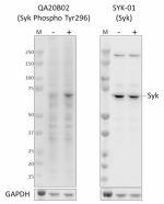
Whole cell extracts (15 µg protein) from serum-starved A20 cells untreated (-) or treated (+) with 5 mM H2O2 for 3 minutes were resolved on a 4-12% Bis-Tris gel, transferred to a PVDF membrane, and probed with 1.0 µg/mL (1:500 dilution) of purified anti-Syk Phospho (Tyr296) recombinant (clone QA20B02) for 2 hours at room temperature. Proteins were visualized by chemiluminescence detection using HRP goat anti-mouse IgG (Cat. No. 405306) at a 1:3000 dilution. Equal Syk loading was confirmed by probing membranes with purified anti-Syk (clone SYK-01) (Cat. No. 626202), at 1.0 µg/mL (1:500 dilution). Direct-Blot™ HRP anti-GAPDH (Cat. No. 607904) was used as a loading control at a 1:50000 dilution (lower). Western-Ready™ ECL Substrate Premium Kit (Cat. No. 426319) was used as a detection agent. Lane M: Molecular weight marker -

Serum starved Ramos cells either untreated (negative control, open histogram) or treated with H2O2 (positive control, filled histogram) were fixed and permeabilized using True-Phos™ Perm Buffer (Cat. No. 425401) and intracellularly stained with Purified anti-Syk Phospho (Tyr296) Recombinant Antibody (clone QA20B02) or Purified Mouse IgG2b, κ Isotype Control (open histogram, dashed line) (representative histogram for either untreated or treated cells) (Cat. No. 402201) followed by Alexa Fluor® 647 Goat anti-mouse IgG (Cat. No. 405322). -

Serum starved Ramos cells treated with H2O2 were fixed and permeabilized using True-Phos™ Perm Buffer Set (Cat. No. 425401). Cell were treated with (negative control, open histogram) or without Lambda Protein Phosphatase (LPP) (positive control, filled histogram) and then intracellularly stained with Purified anti-Syk Phospho (Tyr296) Recombinant Antibody (clone QA20B02) or Purified Mouse IgG2b, κ Isotype Control (open histogram, dashed line) (representative histogram for either untreated or treated cells) (Cat. No. 402201) followed by Alexa Fluor® 647 Goat anti-mouse IgG (Cat. No. 405322).
| Cat # | Size | Price | Quantity Check Availability | ||
|---|---|---|---|---|---|
| 949301 | 25 µg | $129.00 | |||
| 949302 | 100 µg | $323.00 | |||
Syk is a non-receptor tyrosine kinase that is a member of the Syk family of tyrosine kinases, with broad expression in hematopoietic cells. Upon immune receptor engagement, phosphorylated ITAM motifs within the cytosolic region of the activated receptor become phosphorylated and recruit Syk to the activated receptor via SH2 domains. ITAM-bound Syk is catalytically active, transmitting signals to multiple downstream signaling partners that drives processes such as phagocytosis, proliferation, and differentiation.
Recent evidence also suggests a role for Syk in non-immune cells. Elevated Syk activation is associated with B-cell malignancies, rheumatoid arthritis, and Alzheimer’s disease, and multiple Syk inhibitors are being developed in clinical trials. Syk activity and function are both regulated by tyrosine phosphorylation at multiple sites. Tyr323 and Tyr352 phosphorylation facilitate binding to downstream effectors, and phosphorylation of Tyr525/Tyr526 in the activation loop is required for kinase activity. Tyr296 phosphorylation has been reported in lung epithelial cancer cells, and has been shown to occur as part of an autophosphorylation mechanism in vitro. This site is embedded within a larger N-terminal linker region that is required for nuclear localization in breast cancer cells. Despite rapid autophosphorylation of this site upon Syk activation, it is not required for Syk activity.
Product Details
- Verified Reactivity
- Human, Mouse
- Antibody Type
- Recombinant
- Host Species
- Mouse
- Immunogen
- Synthetic peptide corresponding to Syk phosphorylated at tyrosine 296
- Formulation
- Phosphate-buffered solution, pH 7.2, containing 0.09% sodium azide
- Preparation
- The antibody was purified by affinity chromatography.
- Concentration
- 0.5 mg/mL
- Storage & Handling
- The antibody solution should be stored undiluted between 2°C and 8°C.
- Application
-
WB - Quality tested
ICFC - Verified - Recommended Usage
-
Each lot of this antibody is quality control tested by western blotting. For western blotting, the suggested use of this reagent is 0.5 - 1.0 µg/mL. For flow cytometric staining using our True-Phos™ Perm Buffer, the suggested use of this reagent is ≤ 1.0 µg per million cells in 100 µL volume. It is recommended that the reagent be titrated for optimal performance for each application.
- Application Notes
-
When Syk is phosphorylated at tyrosine 296, the protein may display a slightly higher observed molecular weight by western blot.
- RRID
-
AB_2910515 (BioLegend Cat. No. 949301)
AB_2910515 (BioLegend Cat. No. 949302)
Antigen Details
- Structure
- Syk isoform A is a 635 amino acid protein with a predicted molecular weight of 72 kD. Syk isoform B is a 612 amino acid protein with a predicted molecular weight of 69.5 kD. Syk isoform B lacks the Tyr296 site.
- Distribution
-
Plasma membrane
- Function
- B-cell receptor, integrin signaling
- Biology Area
- Cell Biology, Immunology, Signal Transduction
- Molecular Family
- Protein Kinases/Phosphatase
- Antigen References
-
- Mansueto MS, et al. 2019. J Biol Chem. 294:7658-7668.
- Geahlen RL. 2009. Biochim Biophys Acta. 1793:1115-27.
- Taniguchi T, et al. 1991. J Biol Chem. 266:15790-6.
- Toyabe S, et al. 2001. Immunology. 103:164-71.
- Gene ID
- 6850 View all products for this Gene ID
- UniProt
- View information about Syk Phospho on UniProt.org
Other Formats
View All Syk Phospho Reagents Request Custom Conjugation| Description | Clone | Applications |
|---|---|---|
| Purified anti-Syk Phospho (Tyr296) Recombinant Antibody | QA20B02 | WB,ICFC |
Compare Data Across All Formats
This data display is provided for general comparisons between formats.
Your actual data may vary due to variations in samples, target cells, instruments and their settings, staining conditions, and other factors.
If you need assistance with selecting the best format contact our expert technical support team.
-
Purified anti-Syk Phospho (Tyr296) Recombinant Antibody

Whole cell extracts (15 µg protein) from serum-starved Ramos... 
Whole cell extracts (15 µg protein) from serum-starved A20 c... 
Serum starved Ramos cells either untreated (negative control... 
Serum starved Ramos cells treated with H2O2<...
