- Clone
- W19027A (See other available formats)
- Regulatory Status
- RUO
- Other Names
- Tripartite Motif Containing 28; RING Finger Protein 96; Transcription Intermediary Factor1-β, KRAB [Kruppel-Associated Box Domain]-Associated Protein 1; KRAB-Associated Protein 1
- Isotype
- Rat IgG2a, κ

-

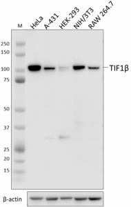
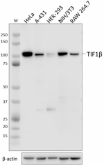
Whole cell extracts (15 µg total protein) from the indicated cell lines were resolved by 4-12% Bis-Tris gel electrophoresis, transferred to a PVDF membrane, and probed with 1.0 µg/mL (1:500 dilution) purified anti-TIF1β (KAP-1, TRIM28) antibody (clone W19027A) overnight at 4°C. Proteins were visualized by chemiluminescence detection using HRP goat anti-rat IgG antibody (Cat. No. 405405) at a 1:3000 dilution. Direct-Blot™ HRP anti-β-actin antibody (Cat. No. 664804) was used as a loading control at a 1:25000 dilution (lower). Lane M: Molecular weight marker. -

HeLa cells were fixed with 4% paraformaldehyde for 10 minutes, permeabilized with methanol for 6 minutes, and blocked with 5% FBS for 60 minutes. Cells were then intracellularly stained with 5.0 µg (1:100 dilution) purified rat IgG2a, κ isotype ctrl antibody (panel A) (Cat. No. 400501) or purified anti-TIF1β (KAP-1, TRIM28) antibody (clone W19027A) (panel B) overnight at 4°C followed by incubation with Alexa Fluor® 594 goat anti-rat IgG antibody (Cat. No. 405422) at 2.0 µg/mL. Nuclei were counterstained with DAPI and the image was captured with a 60X objective. -

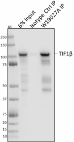
Whole cell extracts (250 µg total protein) prepared from HeLa cells were immunoprecipitated overnight with 2.5 µg purified rat IgG2a, κ isotype ctrl antibody (Cat. No. 400501) or purified anti-TIF1β (KAP-1, TRIM28) antibody (clone W19027A). The resulting IP fractions and whole cell extract input (6%) were resolved by 4-12% Bis-Tris gel electrophoresis, transferred to a PVDF membrane and probed with purified anti-TIF1β (KAP-1, TRIM28) antibody (clone 20A1) (Cat. No. 619301). Lane M: Molecular weight marker.
| Cat # | Size | Price | Quantity Check Availability | ||
|---|---|---|---|---|---|
| 941301 | 25 µg | $118.00 | |||
| 941302 | 100 µg | $293.00 | |||
TIFβ (transcription intermediary factor 1-beta) is an 89 kD member of the tripartite motif family. This protein contains three zinc binding domains, a RING domain, a B-box type 1 and type 2 domain, and a coiled-coil region. TIFβ is found in the nucleus and associates with specific chromatin regions. This protein forms a complex with KRAB-domain transcription factors and recruits SETDB1 to histone 3 to increase KRAB-mediated transcriptional repression. TIF1β has been reported to interact with SETDB1 and CBX3 proteins. Studies using knockout mice reveal the important function of TIF1β in regulating genomic imprinting, T cell activation, and T cell tolerance.
Product Details
- Verified Reactivity
- Human, Mouse
- Antibody Type
- Monoclonal
- Host Species
- Rat
- Immunogen
- Full-length recombinant mouse TIF1β protein
- Formulation
- Phosphate-buffered solution, pH 7.2, containing 0.09% sodium azide
- Preparation
- The antibody was purified by affinity chromatography.
- Concentration
- 0.5 mg/mL
- Storage & Handling
- The antibody solution should be stored undiluted between 2°C and 8°C.
- Application
-
WB - Quality tested
IP, ICC - Verified - Recommended Usage
-
Each lot of this antibody is quality control tested by western blotting. For western blotting, the suggested use of this reagent is 1.0 µg/mL. For immunocytochemistry, a concentration range of 1.0 - 5.0 μg/mL is recommended. For immunoprecipitation, the suggested use of this reagent is 2.5 µg/test. It is recommended that the reagent be titrated for optimal performance for each application.
- Application Notes
-
- BioLegend offers two monoclonal antibodies, clones 20A1 and W19027A that bind TIF1β (KAP-1, TRIM28).
- 20A1 is a mouse IgG1, κ isotype; W19027A is a rat IgG2a, κ isotype
- Both clones detect human and mouse TIF1β by western blot; 20A1 binds TIF1β with a higher affinity than W19027A for this application, but also shows a higher background
- Both clones are validated for immunocytochemistry on human cells
- W19027A is validated for immunoprecipitation, 20A1 has not been tested for this application
- Clone W19027A was tested for immunocytochemistry using 4% PFA-fixed HeLa cells permeabilized with either Triton X-100 or methanol. Both methods were compatible with TIF1β staining. - RRID
-
AB_2888901 (BioLegend Cat. No. 941301)
AB_2888901 (BioLegend Cat. No. 941302)
Antigen Details
- Structure
- TIF1β is an 835 amino acid protein with a predicted molecular weight of 89 kD.
- Distribution
-
Ubiquitously expressed/Nucleus
- Function
- Chromatin remodeling
- Antigen References
-
1. Ryan RF, et al. 1999. Mol. Cell. Biol. 19:4366.
2. Schultz DC, et al. 2002. Genes Dev. 16:919.
3. Moosmann PR, et al. 1996. Nucleic Acids Res. 24:4859.
4. Friedman JR, et al. 1996. Genes Dev. 10:2067.
5. Messerschmidt DM, et al. 2012. Science 335:1499.
6. Chikuma S, et al. 2012. Nat. Immunol. 13:596. - Gene ID
- 10155 View all products for this Gene ID
- UniProt
- View information about TIF1beta on UniProt.org
Other Formats
View All TIF1β Reagents Request Custom Conjugation| Description | Clone | Applications |
|---|---|---|
| Purified anti-TIF1β (KAP-1, TRIM28) Antibody | W19027A | WB,IP,ICC |
Compare Data Across All Formats
This data display is provided for general comparisons between formats.
Your actual data may vary due to variations in samples, target cells, instruments and their settings, staining conditions, and other factors.
If you need assistance with selecting the best format contact our expert technical support team.


