- Regulatory Status
- RUO
- Other Names
- DNAX accessory molecule-1, DNAM-1, CD226, Platelet and T-cell activation antigen 1, PTA1, T lineage specific activation antigen 1 antigen, TLiSA

-

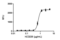
Immobilized human CD226 supports the adhesion of U937 cells in a dose dependent manner with ED50 range of 0.25 - 1.0 µg/mL. Calcein-AM (Cat. No. 425201) was used to measure the number of adherent cells. -
Stability testing for human CD226. Human CD226 was aliquoted in PBS, pH 7.2 at 0.2 mg/ml. One aliquot was freeze and thawed four times (4x freeze/thaws), and compared to a control kept at 4°C (control). The samples were tested in U937 cell adhesion assay.
| Cat # | Size | Price | Quantity Check Availability | ||
|---|---|---|---|---|---|
| 773602 | 10 µg | $83.00 | |||
| 773604 | 25 µg | $311.00 | |||
| 773606 | 100 µg | $604.00 | |||
Select size of product is eligible for a 40% discount! Promotion valid until December 31, 2024. Exclusions apply. To view full promotion terms and conditions or to contact your local BioLegend representative to receive a quote, visit our webpage.
DNAM-1, also known as CD226, was first identified as T lineage-specific activation antigen (TLiSA) and subsequently renamed as Platelet and T-cell activation antigen 1 (PTA). DNAM-1 consists of two Ig-like domains on its extracellular portion, a transmembrane region and a cytoplasmic region containing binding motifs for members of band 4.1 family and membrane-associated guanylate kinase homolog (MAGUK) family. DNAM-1 is predominantly expressed on cell surface of T cells, NK cells, monocytes/ macrophages, platelets and megakaryocytes and a subset of B cells. DNAM-1 serves as a signaling-transducing adhesion molecule, and it physically and functionally associates with LFA-1 (CD11a/CD18) on NK and T cells. The clustering of LFA-1 induces Fyn-mediated phosphorylation of DNAM-1 and signaling. Several evidences demonstrated that DNAM-1 is involved in NK and T cell-mediated cytotoxicity (CTL) against ligand-expressing tumor cells. Two ligands for DNAM-1 have been identified, the poliovirus receptor (PVR/CD155) and its family member nectin-2 (PRR/CD112), which are located at cell junctions and broadly expressed on epithelial, endothelial and neuronal cells. Additionally, DNAM-1 regulates monocyte extravasation through PVR expressed by endothelial cells. It has reported that DNAM-1 is specifically expressed on differentiated Th1 cells, but not to Th2 or Th0 cells. Antibody blocking assay demonstrated that DNAM-1 may play a role in polarization of T cells, activation and effectors of Th1 cells. PVRIG (CD112R) is identified as a novel immune checkpoint to inhibit T cell response by competing DNAM-1 with CD112.
Product Details
- Source
- Human DNAM-1/CD226, amino acid (Glu19-Asn247) (Accession: #Q15762.2), with a linker (GSSR), a C-terminal human IgG1 (Pro100-Lys330) and a 6x His tag, was expressed in 293E cells.
- Molecular Mass
- The 464 amino acid recombinant protein has a predicted molecular mass of approximately 52.3 kD. The DTT-reduced and non-reduced protein migrates at approximately 65 and 130 kD respectively by SDS-PAGE. The predicted N-terminal amino acid is Glu.
- Purity
- > 95%, as determined by Coomassie stained SDS-PAGE.
- Formulation
- 0.22 µm filtered protein solution is in PBS, pH 7.2.
- Endotoxin Level
- Less than 0.1 EU per μg protein as determined by the LAL method.
- Concentration
- 10 and 25 µg sizes are bottled at 200 µg/mL. 100 µg size and larger sizes are lot-specific and bottled at the concentration indicated on the vial. To obtain lot-specific concentration and expiration, please enter the lot number in our Certificate of Analysis online tool.
- Storage & Handling
- Unopened vial can be stored between 2°C and 8°C for up to 2 weeks, at -20°C for up to six months, or at -70°C or colder until the expiration date. For maximum results, quick spin vial prior to opening. The protein can be aliquoted and stored at -20°C or colder. Stock solutions can also be prepared at 50 - 100 µg/mL in appropriate sterile buffer, carrier protein such as 0.2 - 1% BSA or HSA can be added when preparing the stock solution. Aliquots can be stored between 2°C and 8°C for up to one week and stored at -20°C or colder for up to 3 months. Avoid repeated freeze/thaw cycles.
- Activity
- ED50 = 0.25 – 1.0 μg/mL as measured by the ability of immobilized protein to support the adhesion of U937 cells. Calcein-AM (Cat. No. 425201) was used to measure the number of adherent cells.
- Application
-
Bioassay
- Application Notes
-
BioLegend carrier-free recombinant proteins provided in liquid format are shipped on blue-ice. Our comparison testing data indicates that when handled and stored as recommended, the liquid format has equal or better stability and shelf-life compared to commercially available lyophilized proteins after reconstitution. Our liquid proteins are verified in-house to maintain activity after shipping on blue ice and are backed by our 100% satisfaction guarantee. If you have any concerns, contact us at tech@biolegend.com.
-
Application References
(PubMed link indicates BioLegend citation) -
- Fuchs A, Colonna M. 2006. Semin. Cancer Biol. 16: 359.
- Kojima H, et al. 2003. J. Biol. Chem. 278: 36748.
- Burns GF, et al. 1985. J. Exp. Med. 161: 1063.
- Scott JL, et al. 1989. J. Biol. Chem. 264: 13475.
- Shibuya A, et al. 1996. Immunity. 4: 573.
- Shibuya K et al. 1999. Immunity. 11: 615.
- Ralston KJ, et al. 2004. J. Biol. Chem. 279: 33816.
- Tahara-Hanaoka S, et al. 2006. Blood. 107: 1491.
- Bottino C, et al. 2003. J. Exp. Med. 198: 557.
- Reymond N, et al. 2004. J. Exp. Med. 199: 1331.
- Dardalhon V, et al. 2005. J. Immunol. 175: 1558.
- Shibuya K, et al. 2003. J. Exp. Med. 198: 1829.
- Zhu Y, et al. 2016. J. Exp. Med. 213: 167.
Antigen Details
- Structure
- Disulfide bond-linked homodimer, Ig domain containing adhesion molecule.
- Distribution
-
Peripheral blood T cells, NK cells, monocytes/macrophages, platelets and megakaryocytes and a subset of B cells; plasma membrane.
- Function
- It is involved in intercellular adhesion, lymphocyte signaling, cytotoxicity and lymphokine secretion mediated by cytotoxic T-lymphocyte (CTL) and NK cells.
- Interaction
- Epithelial, endothelial and neuronal cells; LFA-1.
- Ligand/Receptor
- PVR (CD155) and Nectin-2 (CD112)
- Bioactivity
- Measured by the ability of immobilized protein to support the adhesion of U937 cells.
- Biology Area
- Immunology
- Molecular Family
- Adhesion Molecules, CD Molecules, Soluble Receptors
- Gene ID
- 10666 View all products for this Gene ID
- UniProt
- View information about DNAM-1 on UniProt.org

