- Regulatory Status
- RUO
- Other Names
- Placental growth factor 1 (PGF1)

-

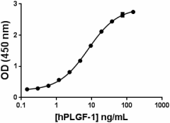
Human PLGF binding to human VEGFR1 by functional ELISA.
| Cat # | Size | Price | Quantity Check Availability | ||
|---|---|---|---|---|---|
| 590702 | 10 µg | $129.00 | |||
| 590704 | 25 µg | $229.00 | |||
| 590706 | 100 µg | $686.00 | |||
Select size of product is eligible for a 40% discount! Promotion valid until December 31, 2024. Exclusions apply. To view full promotion terms and conditions or to contact your local BioLegend representative to receive a quote, visit our webpage.
Placenta growth factor (PLGF) was initially cloned form a term placenta cDNA library. PLGF is a dimeric glycoprotein with 53% homology to VEGF. As a result of alternative splicing, four isoforms for PLGF has been described, PLGF-1, PLGF-2, PLGF-3, and PLGF-4. PLGF-2 binds to heparin with high affinity, whereas neither PLGF-1, PLGF-3, nor PLGF-4 bind heparin. PLGF-2 is the only isoform in mouse. PLGF binds to VEGFRI but not to VEGFRII; also PLGF binds to sVEGF1. PLGF plays a key role in the maternal vascular function during pregnancy. Low levels of PLGF have been associated with patients experiencing preeclampsia and delivering small-for-gestational age (SGA) neonates. High expression of PLGF has been associated to colorectal cancer (CRC), and its expression in CRC tissues correlates with high levels of MMP-9 and VEGF1. PLGF has been shown to increase tumor cell migration in lung cancer, leukemia, and melanoma cells. In colorectal cancer, PLGF induces tumor cell migration and invasion through upregulation of MMP9. PLGF is expressed in the majority of medulloblastomas, and high expression of PLGF receptor neuropilin 1 (Nrp1) correlates with poor survival in patients. In a mouse model, PLGF/Nrp1 blockade provided direct antitumor effects in vivo, medulloblastoma regression, decreased metastasis, and increased survival.
Product Details
- Source
- Human PLGF, amino acids (Ala21-Arg149) (Accession# NM_001207012) was expressed in 293E cells.
- Molecular Mass
- The 129 amino acid recombinant protein has a predicted molecular mass of approximately 14.5 kD. The DTT-reduced and non-reduced protein migrate at approximately 20-25 kD and 45-50 kD by SDS-PAGE respectively. The N-terminal amino acid is Ala.
- Purity
- >95%, as determined by Coomassie stained SDS-PAGE.
- Formulation
- 0.22 µm filtered protein solution is in 5 mM citric acid, 0.15 M NaCl, 5 mM NaH2PO4, pH 4.0.
- Endotoxin Level
- Less than 1.0 EU/µg cytokine as determined by the LAL method.
- Concentration
- 10 and 25 µg sizes are bottled at 100 µg/mL. 100 µg size and larger sizes are lot-specific and bottled at the concentration indicated on the vial. To obtain lot-specific concentration and expiration, please enter the lot number in our Certificate of Analysis online tool.
- Storage & Handling
- Unopened vial can be stored between 2°C and 8°C for up to 2 weeks, at -20°C for up to six months, or at -70°C or colder until the expiration date. For maximum results, quick spin vial prior to opening. The protein can be aliquoted and stored at -20°C or colder. Stock solutions can also be prepared at 50 - 100 µg/mL in appropriate sterile buffer, carrier protein such as 0.2 - 1% BSA or HSA can be added when preparing the stock solution. Aliquots can be stored between 2°C and 8°C for up to one week and stored at -20°C or colder for up to 3 months. Avoid repeated freeze/thaw cycles.
- Activity
- ED50 = 2- 12 ng/ml as determined by the dose dependent binding to VEGFR1-Fc chimera using a functional ELISA.
- Application
-
Bioassay
- Application Notes
-
BioLegend carrier-free recombinant proteins provided in liquid format are shipped on blue-ice. Our comparison testing data indicates that when handled and stored as recommended, the liquid format has equal or better stability and shelf-life compared to commercially available lyophilized proteins after reconstitution. Our liquid proteins are verified in-house to maintain activity after shipping on blue ice and are backed by our 100% satisfaction guarantee. If you have any concerns, contact us at tech@biolegend.com.
Antigen Details
- Structure
- Homodimer
- Distribution
-
Vascular endothelial cells, smooth muscle cells, keratinocytes, hematopoitic cells, retinal pigment epithelial cells, tumor cells, and cerebellar stroma cells.
- Function
- PLGF is an angiogenic factor and induces proliferation and migration of endothelial cells. It stimulates endothelial cells and synergistically amplifies the action of VEGF. Soluble VEGFR1 (sFLT1) can regulate the accessibility of PLGF.
- Interaction
- Endothelial cells, Macrophages, and trophoblats.
- Ligand/Receptor
- VEGFR1 (FLT-1), and neuropilin 1 (Nrp1).
- Cell Type
- Hematopoietic stem and progenitors
- Biology Area
- Cell Biology, Signal Transduction, Stem Cells
- Molecular Family
- Cytokines/Chemokines, Growth Factors
- Antigen References
-
1. Maglione D, et al. 1991. Proc. Natl. Acad. Sci. USA 88:9267.
2. Carmeliet P, et al. 2001. Nat. Med. 7:575.
3. Casalou C, et al. 2007. Leukemia 21:1590.
4. Marcellini M, et al. 2006. Am. J. Pathol. 169:643.
5. Yao J, et al. 2011. Proc. Natl. Acad. Sci. USA 108:11590.
6. Cai J, et al. 2011. PLoS One 6:e18076.
7. Sibiude J, et al. 2012. PLoS One 7:e50208.
8. Snuderl M, et al. 2013 Cell 152:1065. - Gene ID
- 5228 View all products for this Gene ID
- UniProt
- View information about PLGF on UniProt.org
