- Regulatory Status
- RUO
- Other Names
- TGFB, DPD1, transforming growth factor, Transforming Growth Factor Beta 1, TGF-Beta-1

-

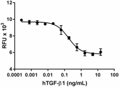
TGF-β1 inhibits the proliferation of mouse HT-2 cells induced by recombinant mouse IL-4 (Cat. No. 574302). The ED50 for this effect is 0.05 – 0.25 ng/mL. -

Stability Testing for Recombinant Human TGF-β1. Recombinant Human TGF-β1 was aliquoted in <30% Acetonitrile, 0.1% TFA (trifluoroacetic acid) at 0.2 mg/mL. One aliquot was frozen and thawed four times (4x Freeze/Thaws) and compared to the control that was kept at 4°C (Control). The samples were tested for their ability to inhibit the proliferation of mouse HT-2 cells induced by recombinant mouse IL-4 (Cat. No. 574302). The ED50 for this effect is 0.05 – 0.25 ng/mL.
| Cat # | Size | Price | Quantity Check Availability | ||
|---|---|---|---|---|---|
| 781802 | 10 µg | $223.00 | |||
| 781804 | 25 µg | $440.00 | |||
TGF-β1 is synthesized in cells as a 390-amino acid. Furin cleaves the protein at residue 278, yielding an N-terminal cleavage product which corresponds to the latency-associated peptide (LAP), and the 25-kD C-terminal portion of the precursor constitutes the mature TGF-β1. TGF-β activators can release TGF-β from LAP. These activators include proteases that degrade LAP, thrombospondin-1, reactive oxygen species, and integrins avb6 and avb8. Mouse TGF-β converts naïve T cells into regulatory T (Treg) cells that prevent autoimmunity. Although human TGF-β1 is widely used for inducing FOXP3+ in vitro, it might not be an essential factor for human Treg differentiation. Th17 murine can be induced from naïve CD4+ T cells by the combination of TGF-β1 and IL-6 or IL-21. Nevertheless, the regulation of human Th17 differentiation is distinct. TGF-β1 seems to have dual effects on human Th17 differentiation in a dose-dependent manner. While TGF-β1 is required for the expression of RORγt, in human naive CD4+ T cells from cord blood, TGF-β1 can inhibit the function of RORγt at high doses. By using serum-free medium, it has been clarified that the optimum conditions for human Th17 differentiation are TGF-β1, IL-1β, and IL-2 in combination with IL-6, IL-21 or IL-23.
Product Details
- Source
- Human TGF-β1, amino acid Ala279 –Ser390 (Accession # P01137) was expressed in CHO cells.
- Molecular Mass
- The 112 amino acid recombinant protein has a predicted molecular mass of approximately 12.7 kD. The DTT-reduced and non-reduced proteins migrate at approximately 14 kD and 28 kD respectively by SDS-PAGE. The predicted N-terminal amino acid is Ala.
- Purity
- >95%, as determined by Coomassie stained SDS-PAGE.
- Formulation
- 0.22 µm filtered protein solution is in 0.1% TFA, 10% Acetonitrile
- Endotoxin Level
- Less than 0.1 EU per µg protein as determined by the LAL method.
- Concentration
- 10 and 25 µg sizes are bottled at 200 µg/mL. 100 µg size and larger sizes are lot-specific and bottled at the concentration indicated on the vial. To obtain lot-specific concentration and expiration, please enter the lot number in our Certificate of Analysis online tool.
- Storage & Handling
- Unopened vial can be stored between 2°C and 8°C for up to 2 weeks, at -20°C for up to six months, or at -70°C or colder until the expiration date. For maximum results, quick spin vial prior to opening. The protein can be aliquoted and stored at -20°C or colder. Stock solutions can also be prepared at 50 - 100 µg/mL in appropriate sterile buffer, carrier protein such as 0.2 - 1% BSA or HSA can be added when preparing the stock solution. Aliquots can be stored between 2°C and 8°C for up to one week and stored at -20°C or colder for up to 3 months. Avoid repeated freeze/thaw cycles.
- Activity
- Human TGF-β1 inhibits the proliferation of mouse HT-2 cells induced by recombinant mouse IL-4 (Cat. No. 574302) The ED50 for this effect is 0.05 – 0.25 ng/mL.
- Application
-
Bioassay
- Application Notes
-
BioLegend carrier-free recombinant proteins provided in liquid format are shipped on blue ice. Our comparison testing data indicates that when handled and stored as recommended, the liquid format has equal or better stability and shelf-life compared to commercially available lyophilized proteins after reconstitution. Our liquid proteins are verified in-house to maintain activity after shipping on blue ice and are backed by our 100% satisfaction guarantee. If you have any concerns, contact us at tech@biolegend.com.
- Product Citations
-
Antigen Details
- Structure
- Dimer
- Distribution
-
TGF-β1 is secreted by numerous cells.
- Function
- TGF-β1 is a multifunctional cytokine that plays pivotal roles in diverse biological processes, including the regulation of cell growth and survival, cell and tissue differentiation, development, inflammation, immunity, hematopoiesis, and tissue remodeling and repair. TGF-β1 is essential for wound healing, stimulates matrix molecule deposition and angiogenesis, and is an essential mediator of the pathologic scarring in fibrotic disorders.
- Interaction
- TGF-beta binding protein (LTBP), epithelial cells, fibroblasts, T cells, B cells, macrophages, and multiple cells respond to TGF-β1.
- Ligand/Receptor
- TGF-β1 binds to type II and type I serine/threonine kinase receptors, which initiate intracellular signals through activation of Smad proteins.
- Cell Type
- Embryonic Stem Cells, Endothelial cells, Fibroblasts, Mesenchymal cells, Mesenchymal Stem Cells, Neural Stem Cells, Osteoclasts, Th17, Tregs
- Biology Area
- Cardiovascular Biology, Immuno-Oncology, Immunology, Stem Cells
- Molecular Family
- Cytokines/Chemokines
- Antigen References
-
- Sun X, et al. 2013. J Virol. 87:10126.
- Benlhabib H, et al. 2015. J Biol Chem. 290:22409-22422.
- Lin C, et al. 2016. J Exp Med. 213:251-271.
- Chen P, et al. 2016. Sci Rep. 6:33407.
- de la Mare JA, et al. 2017. BMC Cancer. 17:202.
- Du C, et al. 2017. Int J Biochem Cell Biol. 90:17-28.
- Nüchel J, et al. 2018. Autophagy. 14:465.
- Gene ID
- 7040 View all products for this Gene ID
- UniProt
- View information about TGF-beta1 on UniProt.org
