- Regulatory Status
- RUO
- Other Names
- Transforming growth factor-beta 1 (TGF-b1), CED, DPD1, TGFB1

| Cat # | Size | Price | Quantity Check Availability | ||
|---|---|---|---|---|---|
| 580709 | 4 pack | $94.00 | |||
Transforming growth factor beta 1 (TGF-β1) is a member of the transforming growth factor beta superfamily of cytokines. The TGF-β1 precursor contains 390 amino acids with an N-terminal signal peptide of 29 amino acids required for secretion from a cell, a 249 amino acid pro-region (latency associated peptide or LAP), and a 112 amino acid C-terminal region that becomes the active TGF-β1 upon activation.
Both LAP and TGF-β1 exist as homodimers in circulation, but the disulfide linked homodimers of LAP and TGF-β1 remain non-covalently associated, forming the small latent TGF-β complex (SLC, 100 kD). The large latent TGF-β complex (LLC, 235-260 kD) contains a third component, the latent TGF-β binding protein (LTBP), which is linked to LAP by a single disulfide bond. LTBP does not confer latency but is for efficient secretion of the complex to extracellular sites. Free active TGF-β1 can be released (activated) by many factors, including enzymes and low or high pH.
TGF-β1 is nearly 100% conserved across mammalian species. It has diverse biological functions in multiple cellular processes such as regulating proliferation and differentiation of various cell types. TGF-β1 is also an important immunoregulatory cytokine, which is involved in the maintenance of self-tolerance, Th17 differentiation, and T-cell homeostasis.
Product Details
- Source
- Human TGF-β1, amino acids Ala279-Ser390 (Accession# NM_000660) was expressed in CHO cells. Mature TGF-β1 was purified from the conditioned media.
- Molecular Mass
- Recombinant human TGF-β1 exists as a disulfide linked homodimer, consisting of two 112 amino acid monomers, each with a predicted molecular mass of approximately 12.8 kD. The non-reduced protein migrates as a homodimer, at approximately 26 kD by SDS-PAGE. The DTT-reduced protein migrates as a monomer, at approximately 13 kD by SDS-PAGE.
- Purity
- >98%, as determined by Coomassie stained SDS-PAGE prior to lyophilization.
- Formulation
- Lyophilized in sterile-filtered PBS, pH 7.2, containing 1% BSA, 0.09% sodium azide, and protease inhibitors.
- Concentration
- Lot-specific (to obtain lot-specific concentration and expiration, please enter the lot number in our Certificate of Analysis online tool.)
- Storage & Handling
- Unopened vials can be stored between 2°C and 8°C until the expiration date. Prior to use, reconstitute the lyophilized powder with 0.2 mL of PBS containing a carrier protein (e.g., 1% BSA, protease free), pH7.4. Re-cap vial, vortex. Allow the reconstituted standard to sit at room temperature for 15 minutes, vortex again to mix completely. The reconstituted standard stock solution can be aliquoted into polypropylene vials and stored at -70°C for up to one month. Do not re-use diluted standards. Avoid repeated freeze/thaw cycles.
- Application
-
ELISA - Quality tested
- Recommended Usage
-
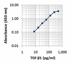
Each lot of this antibody is quality control tested by ELISA assay. A standard curve comprised of two-fold dilutions from 500 pg/ml to 7.8 pg/ml is recommended, and the end-user should titrate each lot of reagents for optimal performance.
- Application Notes
-
To measure TGF-ß1, this recombinant protein can be used as a standard in sandwich ELISA format, when used in conjunction with the purified 21C11 antibody (Cat. No. 525301) as the capture antibody, and the biotinylated 19D8 antibody (Cat. No. 521705) as the detection antibody.
- Product Citations
-
Antigen Details
- Structure
- Homodimer
- Distribution
-
TGF-β1 is secreted by numerous cells.
- Function
- TGF-β1 influences the fate of T cells into different subpopulations depending on the occurrence of other cytokines. TGF-β1 induces growth and survival, cell an tissue differentiation, development, inflammation, immunity, hematopoiesis, tissue remodeling, wound healing, and stimulates ECM deposition and angiogenesis. It is an essential mediator of pathologic scarring in fibrotic disorders.
- Interaction
- Multiple cells express the TGF-β1 receptors.
- Ligand/Receptor
- Heterodimeric receptor consisting of Type I (TbRI) and Type II (TbRII).
- Biology Area
- Apoptosis/Tumor Suppressors/Cell Death, Cell Biology, Immunology, Stem Cells
- Molecular Family
- Cytokines/Chemokines, Growth Factors
- Antigen References
-
1. Dubois CM, et al. 1995. J. Biol. Chem. 270:10618.
2. Dennler S, et al. 1998. EMBO J. 17:3091.
3. Annes JP, et al. 2003. J. Cell. Sci. 116:217.
4. Zou Z and Sun PD. 2004. Protein Expr. Purif. 37:265.
5. Park SH, 2005. J. Biochem. Mol. Biol. 38:9.
6. Valcourt U, et al. 2005. Mol. Biol. Cell. 16:1987.
7. Mucida D, et al. 2007. Science 317:256.
8. Acosta-Rodriguez EV, et al. 2007. Nat. Immunol. 8:942.
9. Puthawala K, et al. 2008. Am. J. Respir. Crit. Care Med. 177:82.
10. Takatori H, et al. 2008. Mod. Rheumatol. 18:533.
11. Manel N, et al. 2008. Nat. Immunol. 9:641.
12. Veldhoen M, et al. 2008. Nat. Immunol. 9:1341.
13. Oida T and Weiner HL. et al. 2011. PLoS ONE 6:e18365. - Gene ID
- 7040 View all products for this Gene ID
- UniProt
- View information about TGF-beta1 on UniProt.org

