- Regulatory Status
- RUO
- Other Names
- CD27 ligand, CD70, Ki-24 antigen, TNFSF7

-

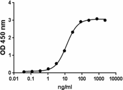
Recombinant mouse CD27L binds to immobilized recombinant mouse CD27 (at 2 µg/mL) in a dose dependent manner. The ED50 = 5 - 30 ng/mL. -
Stability testing for Mouse CD27L: Mouse CD27L was aliquoted in PBS, pH 7 at 0.2 mg/ml and was treated as follow: one aliquot was kept at 4⁰C (control), another was freeze-thawed four times (4x freeze-thaws), one incubated for seven days at room temperature (RT), and another incubated at 37⁰C for one week. After this procedure, the samples were tested for their ability to induce proliferation of mouse T cells when anti-CD3 (1 µg/ml, Cat. No. 100207) is immobilized.
| Cat # | Size | Price | Quantity Check Availability | ||
|---|---|---|---|---|---|
| 550404 | 25 µg | $218.00 | |||
| 550406 | 100 µg | $633.00 | |||
Select size of product is eligible for a 40% discount! Promotion valid until December 31, 2024. Exclusions apply. To view full promotion terms and conditions or to contact your local BioLegend representative to receive a quote, visit our webpage.
CD27L is a 50 kD type II transmembrane glycoprotein and member of the tumor necrosis factor superfamily. Mouse CD27L and its human counterpart share about 56% amino acid sequence identity. The interaction between CD27L and CD27, a protein of the TNF receptor superfamily expressed on NK cells and subsets of T and B cells, provides costimulatory signals that are required for T cell proliferation, clonal expansion, and promotion of effector T cell formation. CD27L-CD27 interaction on mouse B cells on the other hand, has been shown to inhibit terminal differentiation of B cells into plasma cells. CD27L expression is induced by antigen receptor activation in B cells and at low levels in mouse T cells. CD27 interaction with recombinant soluble CD27L protein (sCD27L), has shown that sCD27L enhances IFN-γ secretion by CD56(bright) NK cells in the presence of IL-12, and these NK cells were able to suppress HCV replication. Also, CD8+ T cells stimulated with sCD27L in the presence of Ag showed enhanced proliferation and production of IL-2 and IFN-γ in vitro. In addition, administration of Ag and soluble CD27L induces expansion of Ag-specific CD8+ T cells in vivo. Data suggests that sCD27L is a potent stimulator of antiglioma immune responses that depend on CD8+ T cells; therefore, sCD27L might be an adjuvant for future immunotherapy trials for glioblastoma.
Product Details
- Source
- Mouse CD27L, amino acids (Gln47-Pro195) (Accession# NP_035747.1), was expressed in CHO cells with a N-terminal 9His tag and a linker sequence.
- Molecular Mass
- The 170 amino acid recombinant protein has a predicted molecular mass of approximately 18.6 kD. The protein migrates at about 35 kD in DTT-reducing conditions by SDS-PAGE. The N-terminal amino acid is His.
- Purity
- >90%, as determined by Coomassie stained SDS-PAGE.
- Formulation
- 0.22 µm filtered protein solution is in PBS, pH 7.2.
- Endotoxin Level
- Less than 0.1 EU per µg of protein as determine by the LAL method.
- Concentration
- 25 µg size is bottled at 200 µg/mL. 100 µg size and larger sizes are lot-specific and bottled at the concentration indicated on the vial. To obtain lot-specific concentration and expiration, please enter the lot number in our Certificate of Analysis online tool.
- Storage & Handling
- Unopened vial can be stored between 2°C and 8°C for up to 2 weeks, at -20°C for up to six months, or at -70°C or colder until the expiration date. For maximum results, quick spin vial prior to opening. The protein can be aliquoted and stored at -20°C or colder. Stock solutions can also be prepared at 50 - 100 µg/mL in appropriate sterile buffer, carrier protein such as 0.2 - 1% BSA or HSA can be added when preparing the stock solution. Aliquots can be stored between 2°C and 8°C for up to one week and stored at -20°C or colder for up to 3 months. Avoid repeated freeze/thaw cycles.
- Activity
- Recombinant mouse CD27L binds to immobilized recombinant mouse CD27 (at 2 µg/mL) in a dose dependent manner. The ED50 is 5.0 - 30 ng/mL.
- Application
-
Bioassay
- Application Notes
-
BioLegend carrier-free recombinant proteins provided in liquid format are shipped on blue-ice. Our comparison testing data indicates that when handled and stored as recommended, the liquid format has equal or better stability and shelf-life compared to commercially available lyophilized proteins after reconstitution. Our liquid proteins are verified in-house to maintain activity after shipping on blue ice and are backed by our 100% satisfaction guarantee. If you have any concerns, contact us at tech@biolegend.com.
Antigen Details
- Structure
- Homotrimer
- Distribution
-
Activated T cells, B cells, NK cells, antigen presenting cells, activated plasmacytoid dendritic cells (pDCs), chronic B cell lymphocytic leukemia, and large B cell lymphomas.
- Function
- Role in T cell proliferation, generation of cytolytic and memory T cells, B cell proliferation and differentiation. CD27L is induced by antigen receptor activation in B cells and downregulated by interaction with its receptor CD27.
- Interaction
- T cells, B cells, NK cells.
- Ligand/Receptor
- CD27
- Bioactivity
- Mouse CD27L induces proliferation of mouse T cells.
- Biology Area
- Cancer Biomarkers, Cell Proliferation and Viability, Immunology
- Molecular Family
- Cytokines/Chemokines, Growth Factors, Immune Checkpoint Receptors, Soluble Receptors
- Antigen References
-
1. Bowman MR, et al. 1994. J. Immunol. 152:1756.
2. Shaw J, et al. 2010. Blood 115:3051.
3. Keller AM, et al. 2009. Blood 113:5167.
4. Tesselaar K, et al. 1997. J. Immunol. 159:4959.
5. Akiba H, et al. 1999. J. Immunol. 162:7058.
6. Miller J, et al. 2010. J. Neurosurg. 113:280.
7. Kuka M, et al. 2013. J. Immunol. 191:2282.
8. Jang YS, et al. 2013. Clin. Immunol. 149:379. - Gene ID
- 21948 View all products for this Gene ID
- UniProt
- View information about CD27L on UniProt.org

