- Regulatory Status
- RUO
- Other Names
- CD54, Ly-47, MALA-2, Intercellular adhesion molecule 1 (ICAM-1)

-

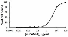
Retinoic acid activated HL60 cells are able to bind to immobilized mouse ICAM-1-Fc chimera in a dose dependent manner. Over 80% of the cells were able to bind mouse ICAM-1 at a dose greater than 12.5 µg/ml (100 µl/well).
| Cat # | Size | Price | Quantity Check Availability | ||
|---|---|---|---|---|---|
| 553004 | 25 µg | $147.00 | |||
| 553006 | 100 µg | $347.00 | |||
ICAM-1, intercellular adhesion molecule 1, is a member of the immunoglobulin superfamily. Structurely, ICAM-1 is characterized by heavy glycosylation with an extracellular domain composed of multiple loops created by disulfide bridges within the protein. The dominant secondary structure of the protein is the beta sheet, leading investigators to hypothesize the presence of dimerization domains within ICAM-1. This protein is constitutively expressed at low levels on the cell surface of leukocytes, endothelial cells, macrophages, and lymphocytes. Upon cytokine activation, by cytokines such as IL-1 and TNFα, the concentration of ICAM-1 on the cell surface greatly increases. The ligand of ICAM-1 is LFA-1 (leukocyte function associated antigen 1), which is found on all T cells. LFA-1/ICAM-1 interaction has been shown to be important for T cell-T cell interactions, which leads to further T cell differentiation. ICAM-1 was found to play a role in infectious diseases, as it was identified as the binding target for entry of the major group of human rhinoviruses into various cell types and is also known for its affinity for Plasmodium falciparum-infected erythrocytes. These findings indicate that ICAM-1 may participate in the pathogenesis of several infectious agents
Product Details
- Source
- Mouse ICAM-1, amino acids (Gln28 - Asn485) (Accession# NP_034623.1), was expressed in CHO cells with a human IgG1 Fc tag at the C-terminus.
- Molecular Mass
- The 696 amino acid recombinant protein, with a C-terminal human IgG1 Fc tag, has a predicted molecular mass of approximately 77 kD. The protein migrates near 130 kD by SDS-PAGE in DTT-reducing conditions and near 250 kD in non-reducing conditions. The predicted N-terminal amino acid is Gln.
- Purity
- >95%, as determined by Coomassie stained SDS-PAGE.
- Formulation
- 0.22 µm filtered protein solution is in PBS.
- Endotoxin Level
- Less than 1.0 EU per µg of protein as determine by the LAL method.
- Concentration
- 25 µg size is bottled at 100 µg/mL. 100 µg size and larger sizes are lot-specific and bottled at the concentration indicated on the vial. To obtain lot-specific concentration and expiration, please enter the lot number in our Certificate of Analysis online tool.
- Storage & Handling
- Unopened vial can be stored between 2°C and 8°C for up to 2 weeks, at -20°C for up to six months, or at -70°C or colder until the expiration date. For maximum results, quick spin vial prior to opening. The protein can be aliquoted and stored at -20°C or colder. Stock solutions can also be prepared at 50 - 100 µg/mL in appropriate sterile buffer, carrier protein such as 0.2 - 1% BSA or HSA can be added when preparing the stock solution. Aliquots can be stored between 2°C and 8°C for up to one week and stored at -20°C or colder for up to 3 months. Avoid repeated freeze/thaw cycles.
- Activity
- Immobilized mouse ICAM-1-Fc chimera is able to bind retinoic acid (2 µM, 72 hr) activated HL60 cells (5 x 10⁴ /well) in a dose dependent manner. Over 80% of the cells were able to bind mouse ICAM-1 at a dose greater than 12.5 µg/ml.
- Application
-
Bioassay
- Application Notes
-
BioLegend carrier-free recombinant proteins provided in liquid format are shipped on blue-ice. Our comparison testing data indicates that when handled and stored as recommended, the liquid format has equal or better stability and shelf-life compared to commercially available lyophilized proteins after reconstitution. Our liquid proteins are verified in-house to maintain activity after shipping on blue ice and are backed by our 100% satisfaction guarantee. If you have any concerns, contact us at tech@biolegend.com.
- Product Citations
-
Antigen Details
- Structure
- Homodimer
- Distribution
- Antigen presenting cells, endothelial cells.
- Function
- ICAM-1, expressed on antigen presenting cells, is able to engage the LFA-1 on T cells. This interaction helps the activation and differentiation of T cells upon TCR-MHC stimulation. ICAM-1 participates in leukocyte transendothelial migration. It is induced by IL-1 and TNF-alpha;.
- Interaction
- T cells.
- Ligand/Receptor
- LFA-1, MAC-1, fibrinogen.
- Bioactivity
- Mouse ICAM-1 is able to bind retinoic acid-activated HL60 cells.
- Cell Type
- Mesenchymal Stem Cells, Embryonic Stem Cells
- Biology Area
- Cell Biology, Neuroscience, Neuroscience Cell Markers, Cell Adhesion, Stem Cells
- Antigen References
-
1. Bella J, et al. 1998. Proc. Natl. Acad. Sci. 95:4140.
2. Katagiri T, et al. 1996. Blood. 87:4276.
3. Rothlein RD. 1986. J. Immunol. 137:1270.
4. Yang L. et al. 2005. Blood. 106:584.
5. Voraberger G. et al. 1991. J. Immunol. 147:2777.
6. Staunton DE, et al. 1988. Cell 52:925.
7. Greve JM, et al. 1989. Cell 56:839.
8. van Buul JD, et al. 2010. PLoS One 5:e11336. - Gene ID
- 15894 View all products for this Gene ID
- UniProt
- View information about ICAM-1 on UniProt.org
