- Regulatory Status
- RUO
- Other Names
- Interleukin-36 beta, Interleukin-1 family member 8, IL-1F8, Interleukin-1 homolog 2, IL-1H2

-

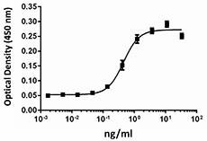
Dose-dependent stimulation of IL-6 secretion in mouse 3T3L1 cells.
| Cat # | Size | Price | Quantity Check Availability | ||
|---|---|---|---|---|---|
| 554502 | 10 µg | $218.00 | |||
| 554506 | 100 µg | $891.00 | |||
| 554508 | 500 µg | $2343.00 | |||
Select size of product is eligible for a 40% discount! Promotion valid until December 31, 2024. Exclusions apply. To view full promotion terms and conditions or to contact your local BioLegend representative to receive a quote, visit our webpage.
The IL-1 family is a group of cytokines comprised of 11 members, including the IL-36 cytokines, IL-36α, β and γ (previously known as IL-1F6, IL-1F8, and IL-1F9). Like other IL-36 cytokines, IL-36β signals through IL-1Rrp2 and IL-1RAcP and activate NF-κB and MAPKs. IL-36β functions as a chemokine and induces migration of bone marrow-derived dendritic cells (BMDCs) and CD4+ T cells. IL-36β induces proinflammatory cytokines, including IL-12, IL-1β, IL-6, and TNF-α in BMDCs. Furthermore, it has been shown that IL-36β synergizes with IL-12 to enhance Th1 polarization. IL-36β is also expressed in neurons and glia cells, but recombinant IL-36β does not induce the classical IL-1 response in brain cells. In a cultured keratinocyte system, IL-36β can induce robust expression of many chemokines in macrophages, T cells, and neutrophils. The secretion of antimicrobial peptides, including HBD-2, HBD-3, lipocalin 2, CAMP, elafin, serpinB1, and IL-8 can be induced by IL-36β in cultured keratinocytes. IL-36β, can also upregulate MMP9 and MMP19 mRNA in cultured keratinocytes. IL-36β has been implicated in the development of human psoriasis. IL-36β is expressed in the skin and the expression can be upregulated by IL-1β, TNF-α, flagellin, and poly (I:C). The expression of IL-36β is also increased in plaque psoriasis. In addition, IL-36β can be detected in mononuclear cells that infiltrate lesional skin. In mouse models of psoriasis, IL-36β is significantly upregulated in inflamed skin.
Product Details
- Source
- Mouse IL-36β, amino acids Ser31-Lys183 (Accession# NM_027163.4), was expressed in E. coli.
- Molecular Mass
- The 153 amino acid recombinant protein has a predicted molecular mass of approximately 17 kD. The DTT-reduced and non-reduced protein migrates at approximately 17 kD by SDS-PAGE. The predicted N-terminal amino acid is Ser.
- Purity
- >95%, as determined by Coomassie stained SDS-PAGE.
- Formulation
- 0.22 µm filtered protein solution is in 20 mM Tris pH7.0, 150 mM NaCl, 2 mM TCEP.
- Endotoxin Level
- Less than 0.01 ng per µg cytokine as determined by the LAL method.
- Concentration
- 10 and 25 µg sizes are bottled at 200 µg/mL. 100 µg size and larger sizes are lot-specific and bottled at the concentration indicated on the vial. To obtain lot-specific concentration and expiration, please enter the lot number in our Certificate of Analysis online tool.
- Storage & Handling
- Unopened vial can be stored between 2°C and 8°C for up to 2 weeks, at -20°C for up to six months, or at -70°C or colder until the expiration date. For maximum results, quick spin vial prior to opening. The protein can be aliquoted and stored at -20°C or colder. Stock solutions can also be prepared at 50 - 100 µg/mL in appropriate sterile buffer, carrier protein such as 0.2 - 1% BSA or HSA can be added when preparing the stock solution. Aliquots can be stored between 2°C and 8°C for up to one week and stored at -20°C or colder for up to 3 months. Avoid repeated freeze/thaw cycles.
- Activity
- The ED50 is 0.1 - 0.5 ng/ml, corresponding to a specific activity 0.2 - 1.0 x 10⁷ units/mg, as determined by a dose-dependent stimulation of IL-6 secretion in 3T3L1 preadipocytes.
- Application
-
Bioassay
- Application Notes
-
BioLegend carrier-free recombinant proteins provided in liquid format are shipped on blue-ice. Our comparison testing data indicates that when handled and stored as recommended, the liquid format has equal or better stability and shelf-life compared to commercially available lyophilized proteins after reconstitution. Our liquid proteins are verified in-house to maintain activity after shipping on blue ice and are backed by our 100% satisfaction guarantee. If you have any concerns, contact us at tech@biolegend.com.
- Product Citations
-
Antigen Details
- Structure
- IL-1 family cytokine.
- Distribution
-
Skin, tonsils, bone marrow, heart, placenta, lung, testis, monocytes, B cells, neurons, and glia cells.
- Function
- IL-36β induces proinflammatory cytokines, synergizes with IL-12 to promote Th1 polarization, and induces antimicrobial peptides. IL-36β is upregulated by IL-1β, TNF-α, flagellin, and poly (I:C); its activity is significantly increased by N-terminal processing.
- Interaction
- Keratinocytes, dendritic cells, macrophages, CD4+ T cells.
- Ligand/Receptor
- Heterodimeric receptor IL-1Rrp2 and IL-1RAcP.
- Biology Area
- Angiogenesis, Cell Biology, Immunology, Innate Immunity, Signal Transduction
- Molecular Family
- Cytokines/Chemokines
- Antigen References
-
1. Towne JE, et al. 2004. J. Biol. Chem. 279:13677.
2. Barksby HE, et al. 2007. Clin. Exp. Immunol. 149:217.
3. Towne JE, et al. 2011. J. Biol. Chem. 286:42594.
4. Wang P, et al. 2005. Cytokine. 29:245.
5. Gresnigt MS and van de Veerdonk FL. 2013. Semin. Immunol. 25:458.
6. Tortola L, et al. 2012. J. Clin. Invest. 122:3965.
7. Johnston A, et al. 2011. J. Immunol. 186:2613. - Gene ID
- 69677 View all products for this Gene ID
- UniProt
- View information about IL-36beta on UniProt.org
