- Regulatory Status
- RUO
- Other Names
- VEGFA, MVCD1, VEGF, Vascular permeability factor (VPF)

-

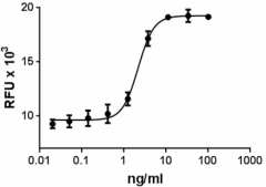
HUVEC cell proliferation induced by mouse VEGF-164.
| Cat # | Size | Price | Quantity Check Availability | ||
|---|---|---|---|---|---|
| 583102 | 10 µg | $218.00 | |||
| 583104 | 25 µg | $382.00 | |||
| 583106 | 100 µg | $703.00 | |||
| 583108 | 500 µg | $1987.00 | |||
VEGF (known also as VEGFA) was initially identified in conditioned medium from bovine pituitary follicular cells. VEGFA belongs to the VEGF family, which has the following members: VEGF-A, VEGF-B, VEGF-C (VEGF-2), VEGF-D, and PlGF (placental growth factor). In addition, viral VEGF homologs (collectively called VEGF-E) and snake venom VEGFs, such as T.f. (Trimeresurus flavoviridis) and svVEGF (called VEGF-F), have been described. VEGFA is alternatively spliced to generate variants with different numbers of amino acids, such as VEGFA120, VEGFA144, VEGFA164, and VEGFA188. VEGFA164 is predominant and responsible for VEGFA biological potency.
While VEGF120 is freely diffusible and does not bind to neuropilins (NRPs) or heparan sulphate (HS), VEGF164 and VEGF187 bind to both, resulting in retention on the cell surface or in the extracellular matrix. NRP1 lacks a typical kinase domain and acts as a co-receptor, and in response to VEGF164, NRP1 couples with VEGF-Rs to signal in endothelial cells. In addition, it has been suggested that bone marrow cells that are recruited to Ewing’s tumors are differentiated into vascular smooth muscle cells, and VEGF164 is responsible for this differentiation.
VEGFA is highly expressed in most of the solid tumors generated in breast, lung, renal, colorectal, and liver tissues. VEGFA has strong vascular permeability activity, and significantly contributes to the formation of ascites tumors. VEGFA can act as a direct proinflammatory mediator during the pathogenesis of rheumatoid arthritis (RA), and protect rheumatoid synoviocytes from apoptosis, which contributes to synovial hyperplasia. VEGFA is expressed in synovial macrophages and synovial fibroblasts in RA patients. Also, VEGFA is associated to age-related macular degeneration (AMD). AMD is due to neovascularization that originates from endothelial cells in the choroid that grow into neurosensory retina as choroidal neovascularization (CNV).
Product Details
- Source
- Mouse VEGF-164, amino acids Ala27-Arg190 (Accession# NM_009505.4) was expressed in 293E cells.
- Molecular Mass
- The 164 amino acid recombinant protein has a predicted molecular mass of approximately 19.27 kD. The DTT-reduced protein migrates at approximately 28 kD and non-reduced protein migrates at 50 kD by SDS-PAGE. The N-terminal amino acid is Alanine.
- Purity
- >98%, as determined by Coomassie stained SDS-PAGE.
- Formulation
- 0.22 µm filtered protein solution is in 5 mM citric acid, 5 mM NaH2PO4, 0.15 M NaCl, pH 4.0.
- Endotoxin Level
- Less than 0.01 ng per µg cytokine as determined by the LAL method.
- Concentration
- 10 and 25 µg sizes are bottled at 200 µg/mL. 100 µg size and larger sizes are lot-specific and bottled at the concentration indicated on the vial. To obtain lot-specific concentration and expiration, please enter the lot number in our Certificate of Analysis online tool.
- Storage & Handling
- Unopened vial can be stored between 2°C and 8°C for up to 2 weeks, at -20°C for up to six months, or at -70°C or colder until the expiration date. For maximum results, quick spin vial prior to opening. The protein can be aliquoted and stored at -20°C or colder. Stock solutions can also be prepared at 50 - 100 µg/mL in appropriate sterile buffer, carrier protein such as 0.2 - 1% BSA or HSA can be added when preparing the stock solution. Aliquots can be stored between 2°C and 8°C for up to one week and stored at -20°C or colder for up to 3 months. Avoid repeated freeze/thaw cycles.
- Activity
- Mouse VEGF-164 induces HUVEC cell proliferation in a dose dependent manner. The ED50 = 0.5 – 3 ng/ml.
- Application
-
Bioassay
- Application Notes
-
BioLegend carrier-free recombinant proteins provided in liquid format are shipped on blue-ice. Our comparison testing data indicates that when handled and stored as recommended, the liquid format has equal or better stability and shelf-life compared to commercially available lyophilized proteins after reconstitution. Our liquid proteins are verified in-house to maintain activity after shipping on blue ice and are backed by our 100% satisfaction guarantee. If you have any concerns, contact us at tech@biolegend.com.
- Product Citations
-
Antigen Details
- Structure
- Homodimer
- Distribution
-
Widely expressed
- Function
- VEGFA is a key player in vasculogenesis, the formation of blood vessels from progenitor cells, as well as angiogenesis. The expression of the VEGFA gene is upregulated via hypoxia, estrogen, and NF-κB pathways. In addition VEGF is upregulated by PDGF-BB, P1GF, TGFβ1, IGF1, FGFs, HGF, TNFα, and IL-1. VEGFA induces proliferation and cell migration in endothelial cells and plays important roles during wound healing. Also, VEGF regulates hematopoietic stem cell survival.
- Interaction
- VEGFA interacts with vascular endothelial cells and monocytes/macrophages, which express VEGFR1. This interaction induces proliferation of endothelial cells and stimulates migration of monocytes/macrophages.
- Ligand/Receptor
- Vascular endothelial growth factor (VEGF)-A binds and activates two tyrosine kinase receptors, VEGFR1 (Flt-1) and VEGFR2 (KDR/Flk-1).
- Cell Type
- Embryonic Stem Cells, Mesenchymal Stem Cells, Neural Stem Cells
- Biology Area
- Angiogenesis, Cell Biology, Neuroscience, Stem Cells, Synaptic Biology
- Molecular Family
- Cytokines/Chemokines, Growth Factors
- Antigen References
-
1. Conn G, et al. 1990 P. Natl. Acad. Sci USA 87:1323.
2. Gerber H, et al. 2002. Nature 417:954.
3. Shibuya M, et al. 2006. J. Biochem. Mol. Biol. 39:469.
4. Shibuya M, et al. 2008. BMB Rep. 41:278.
5. Monaghan-Benson E, et al. 2010. The American Journal of Pathology 177:2091.
6. Koch S and Claesson-Welsh L. 2012. Cold Spring Harb Perspect Med. 2:a006502. - Gene ID
- 22339 View all products for this Gene ID
- UniProt
- View information about VEGF-A on UniProt.org
