- Regulatory Status
- RUO
- Other Names
- Urogastrone (URG), HOMG4, Epidermal Growth Factor

-

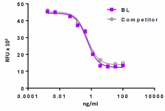
Recombinant rat EGF induces cytotoxic effect on human A431 epidermoid carcinoma cell line in a dose dependent manner. BioLegend’s protein was compared side-by-side to the leading competitor’s equivalent product.
| Cat # | Size | Price | Quantity Check Availability | ||
|---|---|---|---|---|---|
| 558906 | 100 µg | $218.00 | |||
Select size of product is eligible for a 40% discount! Promotion valid until December 31, 2024. Exclusions apply. To view full promotion terms and conditions or to contact your local BioLegend representative to receive a quote, visit our webpage.
Rat EGF was initially isolated from the submaxillary gland of the rat. EGF is a member of the EGF family which comprises of TGF-α, heparin-binding EGF like-growth factor (HB-EGF), epigen, epiregulin, betacellulin, neuroregulin, and tomoregulin. All the EGF family members are synthesized as Type I membrane protein precursors, which can undergo proteolytic cleavage at the plasma membrane to release a mature soluble ectodomain. EGF ligands bind to the ErbB receptors and induce a conformational change in the extracellular domain, which enables homodimer or heterodimer formations that activate the intracellular kinase domain. The EGFR family, ErbB receptors, contains four members that belong to the receptor tyrosine kinase superfamily: EGFR (ErbB1/HER-1), ErbB2 (neu/HER-2), ErbB3 (HER-3), and ErbB4 (HER-4). EGF primarily induces the formation of EGFR/EGFR homodimers and EGFR/ErbB2 heterodimers. EGF is a potent regulator of cell growth and differentiation in multiple tissues. It plays important roles in the regulation of biological processes such as neoplasia, wound healing, gastrointestinal maturation, and nutrient absorption. It is also involved in regulating kidney blood circulation and ion transport. It has been suggested that EGF-ErbB signaling pathways might be key factors in the progress of renal lesions including effects on the regulation of sodium reabsorption in the collecting ducts.
Product Details
- Source
- Rat EGF, amino acids Met-(Asn974-Arg1026) (Accession# NM_012842), was expressed in E. coli.
- Molecular Mass
- The 54 amino acid recombinant protein has a predicted molecular mass of approximately 6.2 kD. The protein migrates at approximately 9 kD in DTT-reducing conditions and at approximately 12 kD in non-reducing conditions by SDS-PAGE. The predicted N-terminal amino acid is Met.
- Purity
- >95%, as determined by Coomassie stained SDS-PAGE.
- Formulation
- 0.22 µm filtered protein solution is in PBS, pH 7.2.
- Endotoxin Level
- Less than 0.01 ng per µg cytokine as determined by the LAL method.
- Concentration
- 10 and 25 µg sizes are bottled at 200 µg/mL. 100 µg size and larger sizes are lot-specific and bottled at the concentration indicated on the vial. To obtain lot-specific concentration and expiration, please enter the lot number in our Certificate of Analysis online tool.
- Storage & Handling
- Unopened vial can be stored between 2°C and 8°C for up to 2 weeks, at -20°C for up to six months, or at -70°C or colder until the expiration date. For maximum results, quick spin vial prior to opening. The protein can be aliquoted and stored at -20°C or colder. Stock solutions can also be prepared at 50 - 100 µg/mL in appropriate sterile buffer, carrier protein such as 0.2 - 1% BSA or HSA can be added when preparing the stock solution. Aliquots can be stored between 2°C and 8°C for up to one week and stored at -20°C or colder for up to 3 months. Avoid repeated freeze/thaw cycles.
- Activity
- ED50 = 0.4 - 2 ng/ml, corresponding to a specific activity of 0.5 - 2.5 x 10⁶ units/mg, as determined by induction of the cytotoxic effect on human A431 epidermoid carcinoma cells.
- Application
-
Bioassay
- Application Notes
-
BioLegend carrier-free recombinant proteins provided in liquid format are shipped on blue-ice. Our comparison testing data indicates that when handled and stored as recommended, the liquid format has equal or better stability and shelf-life compared to commercially available lyophilized proteins after reconstitution. Our liquid proteins are verified in-house to maintain activity after shipping on blue ice and are backed by our 100% satisfaction guarantee. If you have any concerns, contact us at tech@biolegend.com.
Antigen Details
- Structure
- Growth factor.
- Distribution
-
Mammary gland cells, macrophages, gut epithelial cells, cells in the nervous system, and kidney.
- Function
- EGF is a potent regulator of cell growth and differentiation in several tissues. It is induced by NGF in pancreatic B cells.
- Interaction
- Epithelial cells, epidermal cells, and fibroblasts.
- Ligand/Receptor
- EGFR/EGFR homodimer and EGFR/ErbB2 heterodimer.
- Bioactivity
- Induces cytotoxic effect on human A431 epidermoid carcinoma cells.
- Cell Type
- Embryonic Stem Cells, Hematopoietic stem and progenitors, Mesenchymal Stem Cells, Neural Stem Cells
- Biology Area
- Cell Biology, Immunology, Innate Immunity, Neuroscience, Stem Cells, Synaptic Biology
- Molecular Family
- Cytokines/Chemokines, Growth Factors
- Antigen References
-
1. Simpson RJ, et al. 1985. Eur. J. Biochem. 153:629.
2. Gezginci-Oktayoglu S, et al. 2012. Diabetes Metab. Res. Rev. 28:654.
3. Rousselet E, et al. 2011. J. Biol. Chem. 286:9185.
4. Staruschenko A, et al. 2013. Am. J. Physiol. Renal Physiol. 305:F12. - Gene ID
- 25313 View all products for this Gene ID
- UniProt
- View information about EGF on UniProt.org
