- Clone
- LMM741 (See other available formats)
- Regulatory Status
- RUO
- Workshop
- VI MA98
- Other Names
- CSF3R, HG-CSFR, Colony Stimulating Factor 3 receptor
- Isotype
- Mouse IgG1, κ
- Barcode Sequence
- GTTTCCGTCATATAG
| Cat # | Size | Price | Quantity Check Availability | ||
|---|---|---|---|---|---|
| 346111 | 10 µg | $369.00 | |||
CD114 is the receptor of the colony stimulating factor 3 (CSF3, G-CSF), consist of two 130 kD, type I transmembrane chains that form a homodimer. The extracellular domain consists of an immunoglobulin-like domain, a cytokine receptor homologue domain, and three fibronectin type III repeats. CD114 is expressed in all stages of granulocyte differentiation and in monocytes, platelets, endothelial cells, placenta and trophoblasts. The binding of CSF3, results in the activation of many signaling molecules such as Syk, Lyn, Jak1, Jak2, Tyk2, SOCS3, SOCS1, STAT5, and Shp1, resulting in the expression of different target genes that will increase neutrophil precursor survival, proliferation and maturation. In mature neutrophils, it increases survival, superoxide anion generation, arachidonic acid release, production of myeloperoxidase and leukocyte alkaline phosphatase (LAP).
Product Details
- Verified Reactivity
- Human
- Reported Reactivity
- Cynomolgus
- Antibody Type
- Monoclonal
- Host Species
- Mouse
- Immunogen
- CHO cells transfected with hG-CSFR
- Formulation
- Phosphate-buffered solution, pH 7.2, containing 0.09% sodium azide and EDTA
- Preparation
- The antibody was purified by chromatography and conjugated with TotalSeq™-A oligomer under optimal conditions.
- Concentration
- 0.5 mg/mL
- Storage & Handling
- The antibody solution should be stored undiluted between 2°C and 8°C. Do not freeze.
- Application
-
PG - Quality tested
- Recommended Usage
-
Each lot of this antibody is quality control tested by immunofluorescent staining with flow cytometric analysis and the oligomer sequence is confirmed by sequencing. TotalSeq™-A antibodies are compatible with 10x Genomics Single Cell Gene Expression Solutions.
To maximize performance, it is strongly recommended that the reagent be titrated for each application, and that you centrifuge the antibody dilution before adding to the cells at 14,000xg at 2 - 8°C for 10 minutes. Carefully pipette out the liquid avoiding the bottom of the tube and add to the cell suspension. For Proteogenomics analysis, the suggested starting amount of this reagent for titration is ≤ 1.0 µg per million cells in 100 µL volume. Refer to the corresponding TotalSeq™ protocol for specific staining instructions.
Buyer is solely responsible for determining whether Buyer has all intellectual property rights that are necessary for Buyer's intended uses of the BioLegend TotalSeq™ products. For example, for any technology platform Buyer uses with TotalSeq™, it is Buyer's sole responsibility to determine whether it has all necessary third party intellectual property rights to use that platform and TotalSeq™ with that platform. - Additional Product Notes
-
TotalSeq™ reagents are designed to profile protein levels at a single cell level following an optimized protocol similar to the CITE-seq workflow. A compatible single cell device (e.g. 10x Genomics Chromium System and Reagents) and sequencer (e.g. Illumina analyzers) are required. Please contact technical support for more information, or visit biolegend.com/totalseq.
The barcode flanking sequences are CCTTGGCACCCGAGAATTCCA (PCR handle), and BAAAAAAAAAAAAAAAAAAAAAAAAAAAAAA*A*A (capture sequence). B represents either C, G, or T, and * indicates a phosphorothioated bond, to prevent nuclease degradation.
View more applications data for this product in our Scientific Poster Library. -
Application References
(PubMed link indicates BioLegend citation) -
- Layton JE, et al. 1997. Growth Factors. 14:117
- Layton JE, et al. 1999. J. Biol. Chem. 274:17445
- Layton JE, et al. 2001. J. Biol. Chem. 276:36779
- Gupta K, et al. 2014. Blood. 123:2550. PubMed
- RRID
-
AB_2936617 (BioLegend Cat. No. 346111)
Antigen Details
- Structure
- Single chain type I transmembrane molecule of 130 kD. The extracellular domain consists of an immunoglobulin-like domain, a cytokine receptor homologue domain, and three fibronectin type III repeats. To function as a receptor, this molecules form a homod
- Distribution
-
Granulocytes (in all stages of differentiation), monocytes, platelets, endothelial cells, placenta and trophoblasts.
- Function
- Stimulates survival, proliferation, and maturation of neutrophil precursors. In mature neutrophils increases survival, superoxide anion generation, arachidonic acid release, production of myeloperoxidase and leukocyte alkaline phosphatase (LAP).
- Interaction
- Syk, Lyn, Jak1, Jak2, Tyk2, SOCS3, SOCS1, STAT5, Shp1.
- Ligand/Receptor
- CSF3(G-CSF)
- Bioactivity
- Stimulate the proliferation and differentiation of neutrophils precursors and the activation of mature neutrophils.
- Cell Type
- Endothelial cells, Granulocytes, Monocytes, Neutrophils, Platelets
- Biology Area
- Immunology
- Molecular Family
- Adhesion Molecules, CD Molecules, Cytokine/Chemokine Receptors
- Antigen References
-
1. Starnes LM, et al. 2009. Blood 114:1753
2. Skokowa J, et al. 2009. Nat Med. 15:151
3. Ai J, et al. 2008. PLoS One. 3:e3422
4. Germeshausen M, et al. 2008. Curr Opin Hematol. 15:332
5. Marino VJ, Roguin LP, et al. 2008. J Cell Biochem. 103:1512
6. Irandoust MI, et al. 2007. EMBO J. 2:1782 - Gene ID
- 1441 View all products for this Gene ID
- UniProt
- View information about CD114 on UniProt.org
Other Formats
View All CD114 Reagents Request Custom Conjugation| Description | Clone | Applications |
|---|---|---|
| PE anti-human CD114 (G-CSFR) | LMM741 | FC |
| APC anti-human CD114 (G-CSFR) | LMM741 | FC |
| PerCP/Cyanine5.5 anti-human CD114 (G-CSFR) | LMM741 | FC |
| TotalSeq™-A1269 anti-human CD114 (G-CSFR) | LMM741 | PG |
| TotalSeq™-C1269 anti-human CD114 (G-CSFR) | LMM741 | PG |
| TotalSeq™-B1269 anti-human CD114 (G-CSFR) | LMM741 | PG |
Compare Data Across All Formats
This data display is provided for general comparisons between formats.
Your actual data may vary due to variations in samples, target cells, instruments and their settings, staining conditions, and other factors.
If you need assistance with selecting the best format contact our expert technical support team.
-
PE anti-human CD114 (G-CSFR)

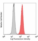
Human peripheral blood granulocytes stained with LMM741 PE -
APC anti-human CD114 (G-CSFR)

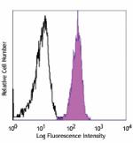
Human peripheral blood granulocytes from stained with LMM741... -
PerCP/Cyanine5.5 anti-human CD114 (G-CSFR)

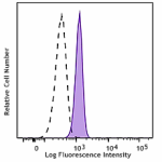
Human peripheral blood granulocytes were stained with CD114 ... -
TotalSeq™-A1269 anti-human CD114 (G-CSFR)
-
TotalSeq™-C1269 anti-human CD114 (G-CSFR)
-
TotalSeq™-B1269 anti-human CD114 (G-CSFR)
