StarBright UltraViolet 575
StarBright™ UltraViolet 575 exhibits peak excitation/emission wavelengths at 340 nm and 569 nm respectively, most closely matching that of BD Horizon™ BUV563. On conventional cytometers, it can be detected with a 355 nm ultraviolet laser and a 586/15 filter set (or equivalent). With proper panel building, it can be used together in panels with StarBright UV740, StarBright UV795, and Spark PLUS UV395™. With its high brightness, StarBright UV575 is best suited for detecting antigens with low to moderate expression levels. In addition, StarBright UV575 can be cocktailed with other fluorophore conjugated antibodies without exhibiting inter-dye interactions. It is also stable towards exposure to heat or a variety of commonly used fixative solutions.
Excitation and Emission Spectra of StarBright UltraViolet 575

Emission spectra (top) and normalized emission spectra (middle) of StarBright UV575 run on a 5-laser Cytek™ Aurora Spectral Cytometer. To compare StarBright UV575 with other fluorophores on a spectral cytometer, use our Aurora Spectral Analyzer tool.
Normalized excitation and emission spectra (bottom) of StarBright UV575 obtained from a spectrophotometer. To compare StarBright UV575 with other fluorophores, use our Fluorescence Spectra Analyzer tool.
Go Beyond BD Horizon™ BUV563

Human peripheral blood lymphocytes were stained with anti-human CD3 (clone UCHT1) Spark Violet™ 423 and anti-human CD4 (clone SK3) conjugated to StarBright UV575 and compared BD Horizon™ BUV563. Samples were acquired on a 5-laser Cytek® Aurora.
Multicolor Staining With StarBright UltraViolet 575
Panel A demonstrates how StarBright UV575 performs in a multicolor panel with fluorophores that share significant spectra overlap with it. Panel B is shown as a reference panel using pre-optimized fluorophores to ensure that staining patterns are similar.

|
Marker
|
Panel A Fluorophore
|
Panel B Fluorophore
|
|
CD4
|
StarBright UV575
|
StarBright UV575
|
|
CD8
|
StarBright UV795
|
Pacific Blue
|
|
CD19
|
Spark PLUS UV395™
|
FITC
|
|
CD56
|
StarBright UV740
|
PE
|
Human PBMCs were stained with the indicated antibodies and analyzed on a spectral flow cytometer. All plots are gated on lymphocytes.
Stability and Validation Testing
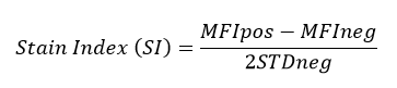
All BioLegend fluorophores undergo rigorous testing procedures to determine how light, heat, and fixation may affect the performance and ensure they will perform reliably. To compare the signal across different conditions and timepoints, we used the Stain Index (formula below) to measure the relative brightness of the antibody.
Photostability Testing
The photostability of StarBright UV575 was tested in two ways that mimic how an antibody may be exposed to light over the course of an experiment.
- Antibodies were stored in the dark or exposed to fluorescent lighting. Then, the antibodies were used to stain freshly harvested cell samples and analyzed immediately.
- Cells were stained with antibody that had been kept under recommended storage conditions. Prior to analysis, the stained cells were stored in the dark or exposed to fluorescent lighting.

To assess the photostability of StarBright UV575-conjugated antibodies, anti-human CD4 (clone SK3) StarBright UV575 was exposed to light or protected in the dark for the indicated times (AB Only). The samples were then used to stain human PBMCs. To measure the photostability of StarBright UV575 stained cells, human PBMCs were stained with anti-human CD4 (clone SK3) StarBright UV575 (+ Cells). Stained cells were exposed to light or kept in the dark as indicated and the staining index of lymphocytes was calculated.
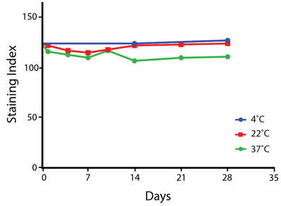
Heat Stability

Anti-human CD4 (clone SK3) StarBright UV575 was aliquoted and incubated at the indicated temperatures over the course of 28 days. The antibodies were then used to stain human lysed whole blood from a single donor and the staining index was calculated.
Fixative Stability
A guide to the fixatives used in this experiment:

Human PBMCs were stained with anti-human CD4 (clone SK3) conjugated to StarBright UV575 and fixed using the respective protocols for each buffer set. Fresh samples were fixed and read on a 5-laser Cytek™ Aurora spectral cytometer immediately. Overnight samples were fixed and stored in Cell Staining Buffer overnight before acquisition. Data shown was gated on lymphocytes.
 Login/Register


Follow Us