- Clone
- SK7; 5.1H11; B73.1; 2D1; SK3; SJ25C1; SK1;
- Regulatory Status
- RUO
- Isotype
- Mouse IgG1, κ (all clones)
- Ave. Rating
- Submit a Review
- Product Citations
- publications

-

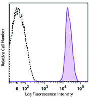
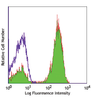
Human peripheral blood was stained with Human TBNK 6 Color Cocktail. Cells were gated on CD45+ (2D1) PerCP/Cyanine5.5 Lymphocytes, and then gated on CD3+ or CD3- (SK7) FITC cell populations. CD3+ cells are shown using CD4 (SK3) PE/Cyanine7 and CD8 (SK1) APC/Fire™ 750. CD3- cells are shown using CD19 (SJ25C1) APC and CD56 (5.1H11) / CD16 (B73.1).
| Cat # | Size | Price | Quantity Check Availability | Save | ||
|---|---|---|---|---|---|---|
| 391503 | 50 tests | 652€ | ||||
This cocktail is intended to differentiate and determine the frequencies of T-Cells, B-Cells, NK-Cells and NKT-Cells in a single test format using either lyse-wash or lyse-no wash human peripheral blood assays. It may be used as a stand-alone product or as a common marker backbone to which additional antibodies may be added for specific applications. The cocktail contains CD3-FITC, CD56-PE, CD16-PE, CD45-PerCP/Cyanine5.5, CD19-APC, CD8-APC/Fire™ 750, CD4-PE/Cyanine7.
CD3 (clone SK7): Is a CD3ε, a 20 kD chain of the CD3/T-cell receptor (TCR) complex found on all mature T lymphocytes, NK-T cells and some thymocytes.
CD45 (clone 2D1): Is a 180 - 240 kD single chain type I membrane glycoprotein also known as leukocyte common antigen (LCA) and T200. It is a tyrosine phosphatase expressed on the plasma membrane of all hematopoietic cells, except erythrocytes or platelets. CD45 is a signaling molecule that regulates a variety of cellular processes including cell growth, differentiation, cell cycle, and oncogenic transformation. CD45 plays a critical role in T and B cell antigen receptor-mediated activation by dephosphorylating substrates including p56Lck, p59Fyn, and other Src family kinases. CD45 non-covalently associates with lymphocyte phosphatase-associated phosphoprotein (LPAP) on T and B lymphocytes. CD45 has been reported to bind galectin-1 and to be associated with several other cell surface antigens including CD1, CD2, CD3, and CD4.
CD56 (clone 5.1H11): Is a single transmembrane glycoprotein also known as NCAM (neural cell adhesion molecule), Leu-19, or NKH1. It is a member of the Ig superfamily. The 140 kD isoform is expressed on NK and NKT cells. CD56 is also expressed in the brain (cerebellum and cortex) and at neuromuscular junctions. Certain large granular lymphocyte (LGL) leukemias, small-cell lung carcinomas, neuronal derived tumors, myelomas, and myeloid leukemias also express CD56. CD56 plays a role in homophilic and heterophilic adhesion via binding to itself or heparan sulfate.
CD16 (clone B73.1): Is known as low affinity IgG receptor III (FcγRIII). It is expressed as two distinct forms (CD16a and CD16b). CD16a (FcγRIIIA) is a 50-65 kD polypeptide-anchored transmembrane protein. It is expressed on the surface of NK cells, activated monocytes, macrophages, a subset of T cells and placental trophoblasts in humans. CD16b (FcγRIIIB) is a 48 kD glycosylphosphatidylinositol (GPI)-anchored protein. Its extracellular domain is over 95% homologous to that of CD16a, and it is expressed specifically on neutrophils. CD16 binds aggregated IgG or IgG-antigen complex which functions in NK cell activation, phagocytosis, and antibody-dependent cell-mediated cytotoxicity (ADCC).
CD19 (clone SJ25C1): Is a 95 kD type I transmembrane glycoprotein also known as B4. It is a member of the immunoglobulin superfamily expressed on B cells (from pro-B to blastoid B cells, absent on plasma cells) and follicular dendritic cells. CD19 is involved in B cell development, activation, and differentiation. CD19 forms a complex with CD21 (CR2) and CD81 (TAPA-1), and functions as a BCR co-receptor.
CD8 (clone SK1): Is a 32-34 kD type I glycoprotein. It forms a homodimer (CD8a/a) or heterodimer (CD8a/b) with CD8b. CD8, also known as T8 and Leu2, is a member of the immunoglobulin superfamily found on the majority of thymocytes, a subset of peripheral blood T cells, and NK cells (which express almost exclusively CD8a homodimers). CD8 acts as a co-receptor with MHC class I-restricted T cell receptors in antigen recognition and T cell activation and has been shown to play a role in thymic differentiation. Two domains in CD8a are important for function: the extracellular IgSF domain binds the α3 domain of MHC class I and the cytoplasmic CXCP motif binds the tyrosine kinase p56 Lck.
CD4 (clone SK3): Also known as T4, is a 55 kD single-chain type I transmembrane glycoprotein expressed on most thymocytes, a subset of T cells, and monocytes/macrophages. CD4, a member of the Ig superfamily, recognizes antigens associated with MHC class II molecules and participates in cell-cell interactions, thymic differentiation, and signal transduction. CD4 acts as a primary receptor for HIV, binding to HIV gp120. CD4 has also been shown to interact with IL-16.
Product Details
- Verified Reactivity
- Human
- Host Species
- Mouse
- Formulation
- Phosphate-buffered solution, pH 7.2, containing 0.09% sodium azide and 0.2% (w/v) BSA (origin USA).
- Preparation
- All antibodies were purified by affinity chromatography and conjugated under optimal conditions.
- Storage & Handling
- The antibody solution should be stored undiluted between 2°C and 8°C, and protected from prolonged exposure to light. Do not freeze.
- Application
-
FC - Quality tested
- Recommended Usage
-
Each lot of this antibody is quality control tested by immunofluorescent staining with flow cytometric analysis. For flow cytometric staining, the suggested use of this reagent is 20 µl per million cells in 100 µl volume. It is recommended that the reagent be titrated for optimal performance for each application.
- RRID
-
AB_2721611 (BioLegend Cat. No. 391503)
Antigen Details
- Distribution
-
T-Cells, B-Cells, NK-Cells and NKT-Cells
- Cell Type
- B cells, NK cells, NKT cells, T cells
- Antigen References
-
SJ25C1:
1. Tedder T, et al. 1994. Immunol. Today 15:437.
2. Bradbury L, et al. 1993. J. Immunol. 151:2915.
SK7:
1. Barclay N, et al. 1993. The Leucocyte FactsBook. Academic Press. San Diego.
2. Beverly P, et al. 1981. Eur. J. Immunol. 11:329.
3. Lanier L, et al. 1986. J. Immunol. 137:2501.
B73.1:
1. Schubert J, et al. 1989. In Leucocyte Typing IV (Knapp W, ed) Oxford University Press Oxford pp 711.
2. Palmer BE, et al. 2005. J. Immunol. 175:8415.
3. Schachner M and Martini R. 1995. Trends Neurosci. 18:183.
4. Wood KL, et al. 2005. Clin. Immunol. 117:294.
5. Björkström NK, et al. 2008. J. Immonol. 181:4219.
5.1H11:
1. Lanier L, et al. 1991. J. Immunol. 146:4421
2. Hemperly J, et al. 1990. J. Mol. Neurosci. 2:71
3. Cremer H, et al. 1994. Nature 367:455.
2D1:
1. Bradstock KF, et al. 1980. J. Natl. Cancer Inst. 65:33.
2. Csiba A, et al. 1984. Br. J. Cancer 50:699.
3. Tchilian EZ, et al. 2001. J. Immunol. 166:1308.
4. Lee MS, et al. 2004. Int. Immunol. 16:1109.SK1:
1. Barclay N, et al. 1993. The Leucocyte Antigen FactsBook. Academic Press Inc. San Diego.SK3:
1. Center D, et al. 1996. Immunol. Today 17:476.
2. Gaubin M, et al. 1996. Eur. J. Clin. Chem. Clin. Biochem. 34:723. - Gene ID
- 925 View all products for this Gene ID 920 View all products for this Gene ID 2215 View all products for this Gene ID 930 View all products for this Gene ID 916 View all products for this Gene ID 4684 View all products for this Gene ID 5788 View all products for this Gene ID 2214 View all products for this Gene ID
Related Pages & Pathways
Pages
Related FAQs
Other Formats
View All Reagents Request Custom Conjugation| Description | Clone | Applications |
|---|---|---|
| Human TBNK 6-Color Cocktail | SK7; 5.1H11; B73.1; 2D1; SK3; SJ25C1; SK1 | FC |
Customers Also Purchased


Compare Data Across All Formats
This data display is provided for general comparisons between formats.
Your actual data may vary due to variations in samples, target cells, instruments and their settings, staining conditions, and other factors.
If you need assistance with selecting the best format contact our expert technical support team.
-
Human TBNK 6-Color Cocktail

Human peripheral blood was stained with Human TBNK 6 Color C...
 Login / Register


Follow Us