 Login / Register


- Clone
- KP1 (See other available formats)
- Regulatory Status
- RUO
- Other Names
- Macrosialin, CD68 antigen, macrophage antigen CD68, scavenger receptor class D, member 1
- Isotype
- Mouse IgG1, κ
- Ave. Rating
- Submit a Review
- Product Citations
- publications

-

IHC staining of anti-CD68 antibody (clone KP1) on formalin-fixed paraffin-embedded Alzheimer´s disease human brain tissue. Following antigen retrieval using Sodium Citrate H.I.E.R., the tissue was incubated with the primary antibody at 5 µg/ml overnight at 4°C. BioLegend's Ultra Streptavidin (USA) HRP Detection Kit (Multi-Species, DAB, Cat. No. 929901) was used for detection followed by hematoxylin counterstaining, according to the protocol provided. Images were captured with a 40X objective. Scale Bar: 20 µm -

IHC staining of anti-CD68 antibody (clone KP1) on formalin-fixed paraffin-embedded Parkinson´s disease human brain tissue. Following antigen retrieval using Sodium Citrate H.I.E.R., the tissue was incubated with the primary antibody at 5 µg/ml overnight at 4°C. BioLegend's Ultra Streptavidin (USA) HRP Detection Kit (Multi-Species, DAB, Cat. No. 929901) was used for detection followed by hematoxylin counterstaining, according to the protocol provided. Images were captured with a 40X objective. -

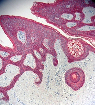
IHC staining of anti-CD68 antibody (clone KP1) on formalin-fixed paraffin-embedded human tonsil tissue. Following antigen retrieval using Retrieve-All Antigen Unmasking System 1 (Cat. No. 927901), the tissue was incubated with the primary antibody at 0.2 µg/ml for 60 minutes at room temperature. BioLegend’s Ultra Streptavidin (USA) HRP Detection Kit (Multi-Species, DAB, Cat. No. 929901) was used for detection followed by hematoxylin counterstaining, according to the protocol provided. -

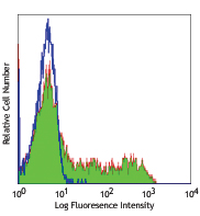
Jurkat cells (negative cell line, panel A) and THP-1 cells (positive cell line, panel B) were fixed with 4% PFA Fixation Buffer (Cat. No. 420801) for 10 minutes and permeabilized with 100% ice-cold methanol for 10 minutes, and blocked with 5% FBS for 1 hour at room temperature. Cells were then stained with Purified anti-CD68 (clone KP1), followed by incubation with Alexa Fluor® 594 Goat anti-mouse IgG (Cat. No. 405326) for 1 hour at room temperature. Nuclei were counterstained with DAPI (Cat. No. 422801). The images were captured on a Revvity Operetta CLS™ High Content Analysis System with a 63X objective and merged. Scale bar: 50 µm
| Cat # | Size | Price | Quantity Check Availability | Save | ||
|---|---|---|---|---|---|---|
| 916104 | 100 µg | 100€ | ||||
Mouse CD68, also known as macrosialin, is an 85-115 kD member of the lysosomal-associated membrane protein (LAMP) family. It is a heavily glycosylated and predominantly intracellular protein, mainly in late endosomes. Macrosialin is the murine homolog to the human macrophage glycoprotein CD68. It is expressed on tissue macrophages, Langerhans cells and at low levels on dendritic cells. High levels of CD68 expression are detected in activated microglia. LAMP proteins may have functions related to cell-cell interaction or cell-ligand interaction. The biological function of CD68 is not completely understood.
Product DetailsProduct Details
- Verified Reactivity
- Human
- Antibody Type
- Monoclonal
- Host Species
- Mouse
- Immunogen
- This antibody was generated using a Lysosomal fraction of human alveolar macrophages.
- Formulation
- Phosphate-buffered solution, pH 7.2, containing 0.09% sodium azide.
- Preparation
- The antibody was purified by affinity chromatography.
- Concentration
- 0.5 mg/ml
- Storage & Handling
- The antibody solution should be stored undiluted between 2°C and 8°C.
- Application
-
IHC-P - Quality tested
ICC - Verified
IP, WB - Reported in the literature, not verified in house - Recommended Usage
-
Each lot of this antibody is quality control tested by formalin-fixed paraffin-embedded immunohistochemical staining. For immunohistochemistry, a concentration range of 0.2 - 5.0 μg/mL is suggested. For immunocytochemistry, a concentration range of 1.25 – 10.0 μg/mL is recommended. It is recommended that the reagent be titrated for optimal performance for each application.
- Application Notes
-
For use in immunocytochemistry (ICC), it is recommended to fix/permeabilize with either of the following:
- Fixation buffer (Cat. No. 420801) with 100% ice-cold methanol
- 100% ice-cold methanol only
Fixation/permeabilization with Fixation buffer (Cat. No. 420801) followed by 0.5 % Triton X-100 is not recommended due to non-specific staining.
- Application References
-
- Holness CL, et al. 1993. Blood 81(6):1607.
- Horny HP, et al. 1990. J. Clin. Path. 43:719.
- Pulford KAF, et al. 1989. J. Clin. Path. 42:414.
- Product Citations
-
- RRID
-
AB_2616797 (BioLegend Cat. No. 916104)
Antigen Details
- Structure
- Sialomucin family, 110 kD.
- Distribution
-
Microglia, monocytes, macrophages, dendritic cells, granulocytes, subset of hematopoietic progenitors, γ/δ T cells, NK cells, LAK cells, subset of B cells, fibroblasts, and endothelial cells.
- Cell Type
- B cells, Dendritic cells, Endothelial cells, Fibroblasts, Granulocytes, Hematopoietic stem and progenitors, Macrophages, Microglia, Monocytes
- Biology Area
- Cell Biology, Immunology, Neuroscience, Neuroscience Cell Markers, Signal Transduction
- Molecular Family
- CD Molecules
- Gene ID
- 968 View all products for this Gene ID
- UniProt
- View information about CD68 on UniProt.org

Other Formats
View All CD68 Reagents Request Custom Conjugation| Description | Clone | Applications |
|---|---|---|
| Purified anti-CD68 | KP1 | IHC-P,ICC,IP,WB |
| Alexa Fluor® 647 anti-CD68 | KP1 | IHC-P,ICC |
| Alexa Fluor® 488 anti-CD68 | KP1 | IHC-P,ICC |
Customers Also Purchased


Compare Data Across All Formats
This data display is provided for general comparisons between formats.
Your actual data may vary due to variations in samples, target cells, instruments and their settings, staining conditions, and other factors.
If you need assistance with selecting the best format contact our expert technical support team.
-
Purified anti-CD68

IHC staining of anti-CD68 antibody (clone KP1) on formalin-f... 
IHC staining of anti-CD68 antibody (clone KP1) on formalin-f... 
IHC staining of anti-CD68 antibody (clone KP1) on formalin-f... 
Jurkat cells (negative cell line, panel A) and THP-1 cells (... -
Alexa Fluor® 647 anti-CD68

IHC staining with Alexa Fluor® 647 anti-CD68 (clone KP1) on ... 
THP-1 cells were fixed and permeabilized with 100% ice-cold ... -
Alexa Fluor® 488 anti-CD68

IHC staining with Alexa Fluor® 488 anti-CD68 (clone KP1) on ... 
Jurkat (negative cell line, panels A and C) and THP-1 cells ...


Follow Us