 Login / Register


- Clone
- W15097A (See other available formats)
- Regulatory Status
- RUO
- Other Names
- Histone deacetylase 3, HD3, SMAP45, RPD3-2
- Isotype
- Mouse IgG2b, κ
- Ave. Rating
- Submit a Review
- Product Citations
- publications

-

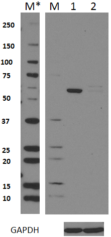
Total lysates (15µg protein) from 293T and 293T/HDAC3 knockdown (KD) cells were resolved by electrophoresis (4-20% Tris-Glycine gel), transferred to nitrocellulose, and probed with 1:500 diluted (1µg/mL) purified anti-HDAC3 antibody, clone W15097A (upper) or 1:3000 diluted anti-GAPDH (poly6314) antibody (lower). Proteins were visualized by chemiluminescence detection using a 1:3000 diluted goat anti-mouse-IgG secondary antibody conjugated to HRP for the anti-HDAC3 antibody, and a donkey anti-rabbit IgG Antibody conjugated to HRP for GAPDH. Lane M: Molecular weight ladder, M* indicates longer exposure. -

Total cell lysate from human HeLa cells (lane 1), human MCF7 cells (lane 2), and mouse NIH-3T3 cells (lane 3) were resolved by electrophoresis, transferred to nitrocellulose, and probed with purified monoclonal anti-HDAC3 (clone W15097A) antibody. Proteins were visualized using an HRP Goat anti-mouse IgG Antibody and chemiluminescence detection. -

HeLa cells were fixed with 4% paraformaldehyde (PFA) for 15 minutes, permeabilized with 0.5% Triton X-100 for three minutes, and blocked with 1% BSA for 60 minutes. Then the cells were intracellularly stained with 2.5 µg/ml purified anti-HDAC3 (clone W15097A) antibody for one hour, followed by staining with Alexa Fluor® 594 conjugated Phalloidin (red) and Alexa Fluor® 488 anti-mouse IgG antibodies (green) for one hour at room temperature. The image was captured with a 40X objective. -

HeLa cells were fixed with 4% paraformaldehyde for 10 minutes, permeabilized with 0.5% Triton X-100 for 10 minutes, and blocked with 5% FBS for 60 minutes. Cells were then intracellularly stained with a 1:100 dilution (5.0 µg/mL) of either Purified Mouse IgG2b, κ Isotype Control Antibody (Cat. No. 402201, panel A) or Purified anti-HDAC3 Antibody, clone W15097A (panel B), overnight at 4° C, followed by incubation with Alexa Fluor® 594 Goat anti-mouse IgG at 2.0 µg/mL (Cat. No. 405326). Nuclei were counterstained with DAPI, and the image was captured with a 60X objective.
| Cat # | Size | Price | Quantity Check Availability | Save | ||
|---|---|---|---|---|---|---|
| 685201 | 25 µg | 100€ | ||||
| 685202 | 100 µg | 231€ | ||||
HDAC3, also known as RPD3-2, HDAC-3, and SMAP45, is a histone deacetylase and is responsible for the deacetylation of lysine residues on core histones and other non-histone substrates. The deacetylation of histones is important for transcriptional regulation, cell cycle progression, and other developmental related functions. HDAC3 represses gene expression by participating in the transcriptional repressor activity of BCL6, by deacetylating H3K27 on enhancer elements. It is also related to participating in transcriptional regulation through binding to YY1, which is a a zinc-finger transcription factor.
Product DetailsProduct Details
- Verified Reactivity
- Human, Mouse
- Antibody Type
- Monoclonal
- Host Species
- Mouse
- Immunogen
- Human HDAC3 recombinant protein (405-424 a.a.) conjugated to KLH.
- Formulation
- Phosphate-buffered solution, pH 7.2, containing 0.09% sodium azide.
- Preparation
- The antibody was purified by affinity chromatography.
- Concentration
- 0.5 mg/mL
- Storage & Handling
- The antibody solution should be stored undiluted between 2°C and 8°C.
- Application
-
WB - Quality tested
KO/KD-WB, ICC - Verified - Recommended Usage
-
Each lot of this antibody is quality control tested by Western blotting. For Western blotting, the suggested use of this reagent is 0.5 - 2.0 µg per mL. For immunocytochemistry, a concentration range of 2.0 - 5.0 μg/mL is recommended. It is recommended that the reagent be titrated for optimal performance for each application.
- Application Notes
-
This clone is not recommended for ChIP (Chromatin Immunoprecipitation) assays (as determined by in-house testing).
- RRID
-
AB_2572203 (BioLegend Cat. No. 685201)
AB_2572203 (BioLegend Cat. No. 685202)
Antigen Details
- Structure
- 428 amino acids with a predicted molecular weight of 49 kD. It belongs to the class I histone deacetylase family.
- Distribution
-
Nucleus.
- Function
- HDAC3 is a deacetylase responsible for the deacetylation of lysine resides on core histones and other non-histone proteins.
- Interaction
- Forms heterodimer with YY1. Interacts with HDAC7, 9, 10, DACH1, DAXX, BCOR, MJD2A/JHDM3A, NRIP1, PRDM6, and SRY.
- Biology Area
- Cell Biology, Chromatin Remodeling/Epigenetics, Neuroscience, Transcription Factors
- Antigen References
-
1. Hua G, et al. 2016. Proc Natl Acad Sci USA. 113:E626.
2. Choi HK, et al. 2015. Nat. Commun. 6:8225.
3. Kim WY, et al. 2015. Int. J. Mol. Med. 35(1):202-10.
4. McHugh CA, et al. 2015. Nature 521:232.
5. Sun Z, et al. 2013. Mol. Cell. 52:769.
6. Sun Z, et al. 2012. Nat. Med. 18:934. - Gene ID
- 8841 View all products for this Gene ID
- UniProt
- View information about HDAC3 on UniProt.org

Related FAQs
Other Formats
View All HDAC3 Reagents Request Custom Conjugation| Description | Clone | Applications |
|---|---|---|
| Purified anti-HDAC3 | W15097A | WB,KO/KD-WB,ICC |
| Alexa Fluor® 594 anti-HDAC3 | W15097A | ICC |
Customers Also Purchased


Compare Data Across All Formats
This data display is provided for general comparisons between formats.
Your actual data may vary due to variations in samples, target cells, instruments and their settings, staining conditions, and other factors.
If you need assistance with selecting the best format contact our expert technical support team.
-
Purified anti-HDAC3

Total lysates (15µg protein) from 293T and 293T/HDAC3 knockd... 
HeLa cells were fixed with 4% paraformaldehyde (PFA) for 15 ... 
Total cell lysate from human HeLa cells (lane 1), human MCF7... 
HeLa cells were fixed with 4% paraformaldehyde for 10 minute... -
Alexa Fluor® 594 anti-HDAC3

HeLa cells were fixed with 4% paraformaldehyde (PFA) for 15 ...


Follow Us