- Clone
- 1A9/S2 (See other available formats)
- Regulatory Status
- RUO
- Other Names
- Spike glycoprotein, S glycoprotein (E2)
- Isotype
- Mouse IgG1, κ
- Ave. Rating
- Submit a Review

| Cat # | Size | Price | Quantity Check Availability | Save | ||
|---|---|---|---|---|---|---|
| 944301 | 25 µg | 137€ | ||||
| 944302 | 100 µg | 324€ | ||||
Severe acute respiratory syndrome coronavirus 2 (SARS-CoV-2) is a single-stranded RNA virus that belongs in a family of viruses known as coronaviruses. SARS-CoV-2 infection, known as COVID-19, was declared a pandemic by WHO on March 11, 2020 and among other symptoms leads to respiratory infection, pulmonary failure which can be fatal. SARS-CoV-2 is structurally composed of 4 main proteins (e.g. spike glycoprotein, envelope glycoprotein, membrane glycoprotein and nucleocapsid protein) and several accessory proteins. The spike glycoprotein (S) is a transmembrane molecule that forms homotrimers on the surface of the virus and facilitates virulence through binding to angiotensin-converting enzyme 2 (ACE2) expressed on host cells. This protein is composed of two subunits, S1 and S2. S1 is responsible for binding to the host cell via ACE2 receptor and the S2 subunit is responsible for fusion with the host cell.
Product DetailsProduct Details
- Verified Reactivity
- SARS-CoV-2
- Antibody Type
- Monoclonal
- Host Species
- Mouse
- Immunogen
- Partial recombinant SARS-CoV protein corresponding to region of S2 subunit
- Formulation
- Phosphate-buffered solution, pH 7.2, containing 0.09% sodium azide
- Preparation
- The antibody was purified by affinity chromatography.
- Concentration
- 0.5 mg/mL
- Storage & Handling
- The antibody solution should be stored undiluted between 2°C and 8°C.
- Application
-
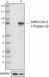
WB - Quality tested
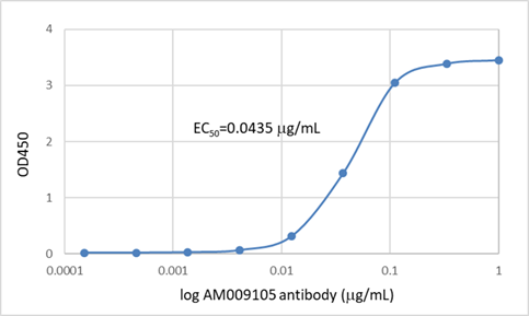
ELISA - Verified
Block - Reported in the literature, not verified in house - Recommended Usage
-
Each lot of this antibody is quality control tested by western blotting. For western blotting, the suggested use of this reagent is 0.0125 - 0.1µg/mL. For ELISA applications, a concentration of 30.4 ng/mL is recommended. It is recommended that the reagent be titrated for optimal performance for each application.
- Application Notes
-
BioLegend offers two clones, 1A9/S2 and A20085C, that recognize SARS-CoV-2 S protein S2.
Both clones have been verified for western blot and direct ELISA, but clone 1A9/S2 is considerably more sensitive for both applications.
Clone 1A9/S2 has been reported to show reactivity towards SARS-CoV (Ng OW, et al. 2014. PLoS One. 9:e102415; Lip KM, et al. 2006. J Virol. 80:941-950.) - Application References
-
- Ng OW, et al. 2014. PLoS One. 9:e102415. (WB, Block)
- Lip KM, et al. 2006. J Virol. 80:941-950. (Block)
- RRID
-
AB_2890871 (BioLegend Cat. No. 944301)
AB_2890871 (BioLegend Cat. No. 944302)
Antigen Details
- Structure
- Spike glycoprotein is a homotrimer. Each monomer consists of 1,273 amino acids with a theoretical molecular weight of 141 kD, and consists of S1 and S2 subunits. The S2 domain is 587 amino acids long with a predicted molecular weight of 64 kD.
- Distribution
-
Viral envelope protein, host cell membrane, host cell endoplasmic reticulum-Golgi intermediate compartment membrane
- Function
- Mediates fusion between virus and host cell membranes
- Interaction
- ACE2
- Ligand/Receptor
- ACE2
- Biology Area
- COVID-19
- Antigen References
-
- Walls AC, et al. 2020. Cell. 181:281-292.
- Yan R, et al. 2020. Science. 367:1444-1448.
- Wrapp D, et al. 2020. Science. 367:1260-1263.
- Shang J, et al. 2020. Proc Natl Acad Sci U S A. 117:11727-11734.
- Gene ID
- 43740568 View all products for this Gene ID
- UniProt
- View information about Viral Protein on UniProt.org
Related Pages & Pathways
Pages
Related FAQs
Other Formats
View All Viral Protein Reagents Request Custom Conjugation| Description | Clone | Applications |
|---|---|---|
| Purified anti-SARS-CoV S Protein S2 | 1A9/S2 | WB,ELISA,Block |
Customers Also Purchased
Compare Data Across All Formats
This data display is provided for general comparisons between formats.
Your actual data may vary due to variations in samples, target cells, instruments and their settings, staining conditions, and other factors.
If you need assistance with selecting the best format contact our expert technical support team.

 Login / Register


Follow Us