- Clone
- 4D10.2 (See other available formats)
- Regulatory Status
- RUO
- Other Names
- Spleen tyrosine kinase, protein tyrosine kinase SYK
- Isotype
- Mouse IgG2a, κ
- Ave. Rating
- Submit a Review
- Product Citations
- publications
-

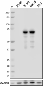
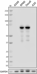
Total cell lysates (15 µg total protein) from A549 (negative control), RPMI-8226, Daudi, and A20 cells were resolved by 4-12% Bis-Tris gel electrophoresis, transferred to a nitrocellulose membrane, and probed with 1.0 µg/mL of Purified anti-Syk Antibody, clone 4D10.2, overnight at 4°C. Proteins were visualized by chemiluminescence detection using HRP goat anti-mouse IgG Antibody (Cat. No. 405306) at a 1:3000 dilution. Direct-Blot™ HRP anti-GAPDH Antibody (Cat. No. 607904) was used as a loading control at a 1:50000 dilution (lower). Lane M: Molecular Weight marker. -
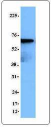
Jurkat cell extract was resolved by electrophoresis, transferred to nitrocellulose and probed with monoclonal anti-Syk (clone 4D10.2) antibody. Proteins were visualized using a goat anti-mouse secondary conjugated to HRP and a chemiluminescence detection system. -

IHC staining using purified anti-Syk (clone 4D10.2) on formalin-fixed paraffin-embedded human tonsil tissue. Following antigen retrieval using 1X Tris-EDTA, pH: 9.0, the tissue was incubated with (B) or without (A) 10 μg/mL of anti-Syk antibody overnight at 4°C, followed by incubation with 2.5 μg/mL of Alexa Fluor® 647 goat anti-mouse IgG (red) (Cat. No. 405322) for one hour at room temperature. Nuclei were counterstained with DAPI (blue) (Cat. No. 422801), and the slide was mounted with ProLong™ Gold Antifade Mountant. The image was captured with a 10x objective. Scalebar = 50 μM -

IHC staining using purified anti-Syk (clone 4D10.2) on formalin-fixed paraffin-embedded human tonsil tissue. The image was captured with a 40x objective. Scalebar = 50 μM
| Cat # | Size | Price | Quantity Check Availability | Save | ||
|---|---|---|---|---|---|---|
| 644301 | 25 µg | 95€ | ||||
| 644302 | 100 µg | 235€ | ||||
Syk is a cytoplasmic 72 kD non-receptor tyrosine kinase that is widely expressed in hematopoietic cells. The Syk kinase associates with a number of proteins including c-Src, Lck, Lyn, Fyn, Vav1, STAT3, SHP1, Grb2, and LAT to couple immunoreceptors to downstream signaling events mediating cellular proliferation, differentiation, and phagocytosis. Syk is modified by phosphorylation on multiple tyrosine sites
Product DetailsProduct Details
- Verified Reactivity
- Human
- Antibody Type
- Monoclonal
- Host Species
- Mouse
- Immunogen
- Human Syk peptide aa314-339
- Formulation
- Phosphate-buffered solution, pH 7.2, containing 0.09% sodium azide.
- Preparation
- The antibody was purified by affinity chromatography.
- Concentration
- 0.5 mg/ml
- Storage & Handling
- The antibody solution should be stored undiluted between 2°C and 8°C.
- Application
-
WB - Quality tested
IHC-P - Verified
IP, ICFC - Reported in the literature, not verified in house - Recommended Usage
-
Each lot of this antibody is quality control tested by Western blotting. Western blotting, suggested working dilution(s): Use 0.5-1 µg per ml antibody dilution buffer. For immunohistochemistry on formalin-fixed paraffin-embedded tissue sections, a concentration range of 1.0 - 10.0 µg/mL is suggested. It is recommended that the reagent be titrated for optimal performance for each application.
- Application Notes
-
This product is for in vitro research use only. It is not to be used for commercial purposes. Use of this product to produce products for sale or for diagnostic therapeutic or drug discovery purposes is prohibited. In order to obtain a license to use this product for commercial purposes, contact The Regents of the University of California.
- Application References
-
- Van Oers, et al. 1995. J. Exp. Med. 182:1585
- Chu DH, et al. 1999. J. Immunol. 163:2610.
- Product Citations
-
- RRID
-
AB_2200267 (BioLegend Cat. No. 644301)
AB_2287062 (BioLegend Cat. No. 644302)
Antigen Details
- Structure
- Non-receptor associated tyrosine kinase, contains 2 SH2 domains, 72 kD
- Distribution
-
Cytoplasmic protein, widely expressed in hematopoietic cells
- Function
- Couples immunoreceptor to downstream signaling events that mediate cellular responses including proliferation, differentiation, and phagocytosis
- Biology Area
- Cell Biology, Immunology, Signal Transduction
- Antigen References
-
1. Taniguchi T, et al. 1991. J. Biol. Chem. 266:15790.
2. Toyabe SL, et al. 2001.Immunology 103:164. - Gene ID
- 6850 View all products for this Gene ID
- UniProt
- View information about Syk on UniProt.org
Related FAQs
Other Formats
View All Syk Reagents Request Custom Conjugation| Description | Clone | Applications |
|---|---|---|
| Purified anti-Syk | 4D10.2 | WB,IP,ICFC,IHC-P |
| PE anti-Syk | 4D10.2 | ICFC |
| APC anti-Syk | 4D10.2 | ICFC |
Compare Data Across All Formats
This data display is provided for general comparisons between formats.
Your actual data may vary due to variations in samples, target cells, instruments and their settings, staining conditions, and other factors.
If you need assistance with selecting the best format contact our expert technical support team.
-
Purified anti-Syk
Total cell lysates (15 µg total protein) from A549 (negative... Jurkat cell extract was resolved by electrophoresis, transfe... 
IHC staining using purified anti-Syk (clone 4D10.2) on forma... 
IHC staining using purified anti-Syk (clone 4D10.2) on form... -
PE anti-Syk

Human peripheral blood lymphocytes were surface stained with... 
Human peripheral blood lymphocytes were surface stained with... 
Human peripheral blood lymphocytes were surface stained with... -
APC anti-Syk
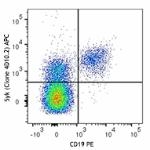
Human peripheral blood lymphocytes were surface stained with... 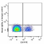
 Login / Register




Follow Us