Purified anti-p63 (ΔN) Antibody

Pricing & Availability
Clone
Poly6190 (See other available formats)
Regulatory Status
RUO
Other Names
p63, δN p63-α, -β, -γ
Isotype
Rabbit Polyclonal IgG
Ave. Rating
Submit a Review
Product Citations
publications
Poly6190_p63-deltaN_092811
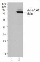
Non-transfected 293 cell lysates (Lane 1) and p63 (deltaN) transfected 293 cell lysates (Lane 2) were resolved by electrophoresis, transferred to nitrocellulose, and probed with anti-p63 (deltaN) antibody (clone Poly6190). Proteins were visualized using a donkey anti-rabbit-IgG secondary conjugated to HRP and chemiluminescence detection.
  • Poly6190_p63-deltaN_092811
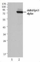
    Non-transfected 293 cell lysates (Lane 1) and p63 (deltaN) transfected 293 cell lysates (Lane 2) were resolved by electrophoresis, transferred to nitrocellulose, and probed with anti-p63 (deltaN) antibody (clone Poly6190). Proteins were visualized using a donkey anti-rabbit-IgG secondary conjugated to HRP and chemiluminescence detection.
Cat # Size Price Quantity Check Availability Save
619001 50 µL 118€
Check Availability


Need larger quantities of this item?
Request Bulk Quote
619002 200 µL 329€
Check Availability


Need larger quantities of this item?
Request Bulk Quote
Description

p63 (ΔN) is a member of the p53 family. This protein has multiple isoforms TA p63-α, -β, γ, ΔN p63-α, -β, and -γ. The molecular weights of the p63 isoforms α=66 kD, β=52 kD, and γ=45 kD. This nuclear protein is essential for limb formation, epidermal morphogenesis, and epithelial stem cell regeneration. ΔN isoforms are antagonistic to p53; TA isoforms can transactivate p53 targets. The p63 ΔN isoforms repress TA isoforms; mutant p53 abrogates p63 transactivation. This protein interacts with p53, p73, and the various TA- and ΔN isoforms. The Poly6190 antibody recognizes human p63 ΔN isoforms and has been shown to be useful for Western blotting.

Product Details
Technical Data Sheet (pdf)

Product Details

Verified Reactivity
Human
Antibody Type
Polyclonal
Host Species
Rabbit
Immunogen
Modified peptide
Formulation
This antibody is provided in phosphate-buffered solution, pH 7.2, containing 0.09% sodium azide and 50% glycerol.
Preparation
The antibody was purified by antigen-affinity chromatography.
Concentration
Lot-specific (to obtain lot-specific concentration and expiration, please enter the lot number in our Certificate of Analysis online tool.)
Storage & Handling
Upon receipt, store frozen at -20°C.
Application

WB - Quality tested
IHC - Reported in the literature, not verified in house

Recommended Usage

Each lot of this antibody is quality control tested by Western blotting. Western blotting, suggested working dilution(s): Use 10 µl per 5 ml antibody dilution buffer for each mini-gel. It is recommended that the reagent be titrated for optimal performance for each application.

Application Notes


This product may contain other non-IgG subtypes.

Additional Product Notes
The 50 µL size can be used for approximately 5 Western blots, and the 200 µL size can be used for approximately 20 Western blots.
Application References
  1. Medawar A, et al. 2008. PLoS ONE 3:e3441. PubMed
  2. Di Girolamo N, et al. 2009. Transplantation. 87:1571. PubMed
  3. Bray LJ, et al. 2012. Biomaterials. 33:3529. PubMed
  4. Zhou Y, et al. 2011. Clin. Invest. Med. 34:E184. (IHC)
  5. Stewart RM, et al. 2015. Invest Ophthalmol Vis Sci. 56:2021. PubMed
  6. Curtis KM, et al. 2015. PLoS One. 10:123642. PubMed
  7. Ahronian LG, et al. 2015. PLoS One.10:123816. PubMed
Product Citations
  1. Marsh T, et al. 2020. Dev Cell. 591:52. PubMed
  2. Hernandez BJ, et al. 2022. Am J Respir Cell Mol Biol. 67:389. PubMed
  3. Hall EG, et al. 2020. Hum Mol Genet. 29:845. PubMed
  4. Goering JP, et al. 2021. Hum Mol Genet. 31:18. PubMed
  5. Ngan SY, et al. 2022. Curr Protoc. 2:e341. PubMed
  6. Teng T, et al. 2022. Development. 149: . PubMed
  7. Saraswathula A, et al. 2023. Int Forum Allergy Rhinol. 13:230. PubMed
  8. Weiner AI 2022. Cell Reports. 41(11):111805. PubMed
  9. Bohnenpoll T, et al. 2016. J Am Soc Nephrol. 2016080849. PubMed
  10. Curtis K, et al. 2015. PLoS One. 10:123642. PubMed
  11. Larsen SB, et al. 2021. Cell Stem Cell. 28:1758. PubMed
  12. Bohnenpoll T, et al. 2017. PLoS Genet.. 10.1371/journal.pgen.1006951. PubMed
  13. Lin-Shiao E, et al. 2018. Genes Dev. 8:9905. PubMed
  14. Zhang B, et al. 2020. Nat Commun. 1.150694444. PubMed
  15. Bray L, et al. 2012. Biomaterials. 33:3529. PubMed
  16. Hazawa M, et al. 2018. EMBO Rep. 0.842361111. PubMed
  17. Wang Z et al. 2018. The Journal of cell biology. 217(6):1941-1955 . PubMed
  18. Li Y, et al. 2017. Mol Cancer Res.. 10.1158/1541-7786.MCR-17-0349. PubMed
  19. Geyer FC, et al. 2018. Nat Commun. 7:2013. PubMed
  20. Mamo TM, et al. 2017. Hum Mol Genet. 26:3553. PubMed
  21. Li M, et al. 2021. Nat Commun. 12:420. PubMed
  22. Traver G, et al. 2017. Free Radic Biol Med. 112:578. PubMed
  23. D'Amico S, et al. 2018. Genes Dev. 32:1175. PubMed
  24. Christians A, et al. 2020. Hum Mol Genet. 1192:29. PubMed
  25. Medawar A, et al. 2008. PLoS One. 2.514583333. PubMed
  26. Kim JE, et al. 2020. Nat Commun. 0.690277778. PubMed
  27. Abbas HA, et al. 2018. Cancer Res. 78:451. PubMed
  28. Zhuang L, et al. 2021. Cells. 10:. PubMed
  29. Rode I, et al. 2015. J Immunol. 195: 5678 - 5687. PubMed
  30. Crowley L, et al. 2020. Elife. 9:00. PubMed
  31. Böttinger P, et al. 2020. Carcinogenesis. 1605:41. PubMed
  32. Lu Y, et al. 2018. Stem Cells. 36:1875. PubMed
  33. Crowell PD et al. 2019. Cell Rep. 28(6):1499-1510 . PubMed
  34. Langa P, et al. 2018. J Dermatol Sci. 89:272. PubMed
  35. Meuser M, et al. 2022. Development. 149:. PubMed
  36. Yuan T, et al. 2020. Front Pharmacol. 11:120. PubMed
  37. Portal C, et al. 2022. Invest Ophthalmol Vis Sci. 63:3. PubMed
  38. Ruiz EJ, et al. 2021. Elife. 10:. PubMed
  39. Wu HH, et al. 2021. Nucleic Acids Res. 49:2740. PubMed
  40. Ahronian L, et al. 2015. PLoS One. 10:123816. PubMed
  41. Chen HJ, et al. 2019. J Exp Med. 216:674. PubMed
  42. Volckaert T et al. 2017. Developmental cell. 43(1):48-59 . PubMed
  43. Aydoğdu N, et al. 2018. Development. 145. PubMed
  44. Stewart R, et al. 2015. Invest Ophthalmol Vis Sci . 56:2021. PubMed
  45. Li M, et al. 2021. Signal Transduct Target Ther. 6:05. PubMed
  46. Masterton S, et al. 2019. Biores Open Access. 8:74. PubMed
  47. Hegde GV et al. 2019. Elife. 8 pii: e46551. PubMed
  48. Van Sciver N, et al. 2021. PLoS Pathog. 17:e1010045. PubMed
  49. Uchtmann KS, et al. 2020. Reprod Toxicol. 91:131. PubMed
  50. Chen M, et al. 2019. Cell Stem Cell. 25:501. PubMed
  51. Assmus AM, et al. 2018. Am J Physiol Renal Physiol. 314:F820. PubMed
  52. Napoli M, et al. 2022. Nat Commun. 13:614. PubMed
  53. Karsli Uzunbas G, et al. 2019. J Biol Chem. 10.1074/jbc.RA119.007965. PubMed
  54. Mao X, et al. 2022. Int J Mol Sci. 23:. PubMed
RRID
SCR_001134 (BioLegend Cat. No. 619001)
SCR_001134 (BioLegend Cat. No. 619002)

Antigen Details

Structure
p53 family, six isoforms, ΔN p63α, β, γ, approximately 66 kD, 52 kD, 45 kD
Distribution

Nuclear

Function
Essential for limb formation, epidermal morphogenesis, epithelial stem cell regeneration. ΔN isoforms antagonistic to p53; TA isoforms can transactivate p53 targets
Interaction
p53, p73, TA- and ΔN isoforms
Biology Area
Apoptosis/Tumor Suppressors/Cell Death, Cell Biology, Cell Cycle/DNA Replication, Transcription Factors
Antigen References

1. Yang A, et al. 1998. Mol Cell. 2:305.
2. Celli J, et al. 1999. Cell. 99:143.
3. Gaiddon C, et al. 2001. Mol Cell Biol. 21:1874.
4. Benard J, et al. 2003. Hum Mutat. 21:182.

Regulation
ΔN isoforms repress TA isoforms, mutant p53 abrogates p63 transactivation
Gene ID
8626 View all products for this Gene ID
UniProt
View information about p63 on UniProt.org
Go To Top Version: 4    Revision Date: 09.22.2023

For Research Use Only. Not for diagnostic or therapeutic use.


This product is supplied subject to the terms and conditions, including the limited license, located at www.biolegend.com/terms) ("Terms") and may be used only as provided in the Terms. Without limiting the foregoing, BioLegend products may not be used for any Commercial Purpose as defined in the Terms, resold in any form, used in manufacturing, or reverse engineered, sequenced, or otherwise studied or used to learn its design or composition without express written approval of BioLegend. Regardless of the information given in this document, user is solely responsible for determining any license requirements necessary for user’s intended use and assumes all risk and liability arising from use of the product. BioLegend is not responsible for patent infringement or any other risks or liabilities whatsoever resulting from the use of its products.


BioLegend, the BioLegend logo, and all other trademarks are property of BioLegend, Inc. or their respective owners, and all rights are reserved.


8999 BioLegend Way, San Diego, CA 92121 www.biolegend.com
Toll-Free Phone: 1-877-Bio-Legend (246-5343) Phone: (858) 768-5800 Fax: (877) 455-9587

This data display is provided for general comparisons between formats.
Your actual data may vary due to variations in samples, target cells, instruments and their settings, staining conditions, and other factors.
If you need assistance with selecting the best format contact our expert technical support team.

ProductsHere

Login / Register
Remember me
Forgot your password? Reset password?
Create an Account