- Clone
- A17080A (See other available formats)
- Regulatory Status
- RUO
- Other Names
- SYN, Synapsin, Synapsin I, Synapsin II, Synapsin III
- Isotype
- Mouse IgG2b, κ
- Ave. Rating
- Submit a Review
- Product Citations
- publications
-

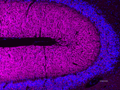
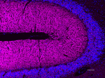
IHC staining of Alexa Fluor® 647 anti-Synapsin I/II/III antibody (clone A17080A) on formalin-fixed paraffin-embedded human cerebellum tissue. Following antigen retrieval using Sodium Citrate H.I.E.R., the tissue was incubated with 2 µg/ml of the primary antibody overnight at 4°C. The slide was mounted with Fluoromount-G with DAPI. The image was captured with a 10X objective. Scale Bar: 100 µm -

IHC staining of Alexa Fluor® 647 anti-Synapsin I/II/III antibody (clone A17080A) on formalin-fixed paraffin-embedded rat cerebellum tissue. Following antigen retrieval using Sodium Citrate H.I.E.R., the tissue was incubated with 2 µg/ml of the primary antibody overnight at 4°C. The slide was mounted with Fluoromount-G with DAPI. The image was captured with a 10X objective. Scale Bar: 100 µm
| Cat # | Size | Price | Quantity Check Availability | Save | ||
|---|---|---|---|---|---|---|
| 853709 | 25 µg | 171 CHF | ||||
| 853710 | 100 µg | 352 CHF | ||||
Synapsins are a family of proteins involved in neurotransmitter release at the chemical synapses. Synapsins anchor synaptic vesicles to the cytoskeletal structures at the presynaptic terminal. Upon receiving an action potential, phosphorylation of synapsins, possibly by PKA, leads to the release of synaptic vesicles from cytoskeletal structures. This allows membrane docking and fusion of synaptic vesicles with the presynaptic membrane and neurotransmitter release.
Product DetailsProduct Details
- Verified Reactivity
- Human, Rat
- Antibody Type
- Monoclonal
- Host Species
- Mouse
- Immunogen
- Recombinant protein corresponding to amino acids 362-450 of human Synapsin I
- Formulation
- Phosphate-buffered solution, pH 7.2, containing 0.09% sodium azide.
- Preparation
- The antibody was purified by affinity chromatography and conjugated with Alexa Fluor® 647 under optimal conditions.
- Concentration
- 0.5 mg/ml
- Storage & Handling
- The antibody solution should be stored undiluted between 2°C and 8°C, and protected from prolonged exposure to light. Do not freeze.
- Application
-
IHC-P - Quality tested
- Recommended Usage
-
Each lot of this antibody is quality control tested by formalin-fixed paraffin-embedded immunohistochemical staining. For immunohistochemistry, a concentration range of 0.5 - 2.0 µg/ml is suggested. It is recommended that the reagent be titrated for optimal performance for each application.
* Alexa Fluor® 647 has a maximum emission of 668 nm when it is excited at 633 nm / 635 nm.
Alexa Fluor® and Pacific Blue™ are trademarks of Life Technologies Corporation.
View full statement regarding label licenses - Excitation Laser
-
Red Laser (633 nm)
- RRID
-
AB_2814566 (BioLegend Cat. No. 853709)
AB_2814566 (BioLegend Cat. No. 853710)
Antigen Details
- Structure
- Synapsin I is a 704 amino acid protein with an apparent molecular mass of ~74 kD. Synapsin II is a 582 amino acid protein with an apparent molecular mass of ~63 kD. Synapsin III is a 580 amino acid protein with an apparent molecular mass of ~63 kD.
- Distribution
-
Tissue distribution: Mainly in the central nervous system. Also expressed in testis.
Cellular distribution: Cytosol, nucleus, and plasma membrane. - Function
- Synapsins are involved in the regulation of synaptic vesicle fusion and neurotransmitter release at the presynaptic terminals.
- Interaction
- CAPON (Carboxyl-terminal PDZ ligand of neuronal nitric oxide synthase protein), PKA
- Cell Type
- Neurons
- Biology Area
- Cell Biology, Neurodegeneration, Neuroinflammation, Neuroscience, Neuroscience Cell Markers, Synaptic Biology
- Molecular Family
- Presynaptic proteins, Synaptic Vesicle Trafficking/Endocytosis
- Antigen References
-
- Greengard P, et al. 1993. Science. 259(5096):780
- Milovanovic D, et al. 2017. Neuron. 93(5):995
- Evergren E, et al. 2007. J. Neurosci. Res. 85(12):2648
- Gene ID
- 6853 View all products for this Gene ID 6854 View all products for this Gene ID 8224 View all products for this Gene ID
- UniProt
- View information about Synapsin I/II/III on UniProt.org
Related FAQs
Other Formats
View All Synapsin I/II/III Reagents Request Custom Conjugation| Description | Clone | Applications |
|---|---|---|
| Purified anti-Synapsin I/II/III | A17080A | WB,IHC-P |
| HRP anti-Synapsin I/II/III | A17080A | WB |
| Biotin anti-Synapsin I/II/III | A17080A | WB,IHC-P |
| Alexa Fluor® 594 anti-Synapsin I/II/III | A17080A | IHC-P |
| Alexa Fluor® 488 anti-Synapsin I/II/III | A17080A | IHC-P |
| Alexa Fluor® 647 anti-Synapsin I/II/III | A17080A | IHC-P |
| Spark YG™ 570 anti-Synapsin I/II/III Antibody | A17080A | IHC-P |
Compare Data Across All Formats
This data display is provided for general comparisons between formats.
Your actual data may vary due to variations in samples, target cells, instruments and their settings, staining conditions, and other factors.
If you need assistance with selecting the best format contact our expert technical support team.
-
Purified anti-Synapsin I/II/III
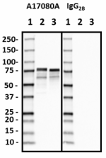
Western blot of purified anti-Synapsin I/II/III antibody (cl... 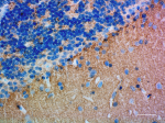
IHC staining of purified anti-Synapsin I/II/III antibody (cl... 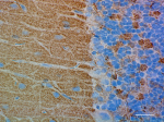
IHC staining of purified anti-Synapsin I/II/III antibody (cl... -
HRP anti-Synapsin I/II/III
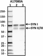
Western blot of HRP anti-Synapsin I/II/III antibody (clone A... -
Biotin anti-Synapsin I/II/III
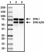
Western blot of Biotin anti-Synapsin I/II/III antibody (clon... 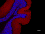
IHC staining of Biotin anti-Synapsin I/II/III antibody (clon... 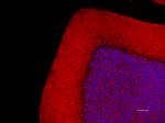
IHC staining of Biotin anti-Synapsin I/II/III antibody (clon... -
Alexa Fluor® 594 anti-Synapsin I/II/III
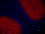
IHC staining of Alexa Fluor® 594 anti-Synapsin I/II/III anti... 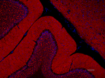
IHC staining of Alexa Fluor® 594 anti-Synapsin I/II/III anti... -
Alexa Fluor® 488 anti-Synapsin I/II/III
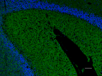
IHC staining of Alexa Fluor® 488 anti-Synapsin I/II/III anti... 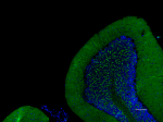
IHC staining of Alexa Fluor® 488 anti-Synapsin I/II/III anti... -
Alexa Fluor® 647 anti-Synapsin I/II/III
IHC staining of Alexa Fluor® 647 anti-Synapsin I/II/III anti... 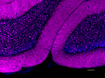
IHC staining of Alexa Fluor® 647 anti-Synapsin I/II/III anti... -
Spark YG™ 570 anti-Synapsin I/II/III Antibody
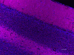
IHC staining of Spark YG™ 570 anti-Synapsin I/II/III antibod... 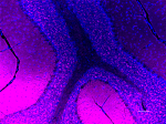
IHC staining of Spark YG™ 570 anti-Synapsin I/II/III antibod...
 Login / Register


Follow Us