- Clone
- LB509 (See other available formats)
- Regulatory Status
- RUO
- Other Names
- NACP, PARK1, PARK4, PD1, Synuclein alpha-140, non-A4 component of amyloid, alpha-synuclein, isoform NACP140, non-A beta component of AD amyloid Parkinson disease (autosomal dominant, Lewy body) 4
- Isotype
- Mouse IgG1, κ
- Ave. Rating
- Submit a Review
- Product Citations
- publications
-

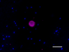
IHC staining of Biotin anti-anti-α-Synuclein, 115-121 antibody (clone LB509) on formalin-fixed paraffin-embedded Parkinson disease brain tissue. Following antigen retrieval using Sodium Citrate H.I.E.R. (10 mM, pH 6.0), the tissue was incubated with 2 µg/mL of the primary antibody overnight at 4°C, followed by incubation with Alexa Fluor® 647 Streptavidin (Cat No. 405237) for one hour at room temperature. Nuclei were counterstained with DAPI. The image was captured with a 40X objective. Scale bar: 50 µm -

IHC staining of Biotin anti-anti-α-Synuclein, 115-121 antibody (clone LB509) on formalin-fixed paraffin-embedded Parkinson disease brain tissue. Following antigen retrieval using Sodium Citrate H.I.E.R. (10 mM, pH 6.0), the tissue was incubated with 2 µg/mL of the primary antibody overnight at 4°C, followed by incubation with Alexa Fluor® 647 Streptavidin (Cat No. 405237) for one hour at room temperature. Nuclei were counterstained with DAPI. The image was captured with a 40X objective. Scale bar: 50 µm
| Cat # | Size | Price | Quantity Check Availability | Save | ||
|---|---|---|---|---|---|---|
| 807709 | 25 µg | 136 CHF | ||||
| 807710 | 100 µg | 311 CHF | ||||
α-synuclein, Alpha-synuclein, is expressed principally in the central nervous system (brain) but is also expressed in low concentrations in a variety of tissues except liver. It is predominantly expressed in the neocortex, hippocampus, substantia nigra, thalamus, and cerebellum of the CNS. It is primarily a neuronal protein, but can also be found in the neuroglial cells. It is concentrated in presynaptic nerve terminals of neurons, as well as having reported nuclear and mitochondrial localization. α-synuclein interacts with plasma membrane phospholipids. α-synuclein in solution is considered to be an intrinsically disordered protein and thus lacks a stable secondary or tertiary structure. However, recent data suggests the presence of partial alpha helical as well as beta sheet structures as well as mostly structured tetrameric states in solution, the equilibrium of which may be altered by binding partners. The human α-synuclein protein is made of 140 amino acids, encoded by the SNCA gene. The primary structure is divided in three distinct domains: (1-60) - An amphipathic N-terminal region dominated by four 11-residue repeats including the consensus sequence KTKEGV. This sequence has a structural alpha helix propensity similar to apolipoproteins-binding domains. (61-95)- a central hydrophobic region which includes the non-amyloid-β component (NAC) region, involved in protein aggregation. (96-140)- a highly acidic and proline-rich region. At least three isoforms of synuclein are produced through alternative splicing. The most common form of the protein, is the full 140 amino acid-long transcript. Other isoforms are alpha-synuclein-126, lacking residues 41-54; and α-synuclein-112, which lacks residues 103-130. α-synuclein may be involved in the regulation of dopamine release and transport and also may function to induce fibrillization of microtubule-associated protein tau. α-synuclein functions as a molecular chaperone in the formation of SNARE complexes. In particular, it can bind to phospholipids of the plasma membrane and to synaptobrevin-2 via its C-terminus domain to influence synaptic activity. α-synuclein is essential for normal development of the cognitive functions and that it significantly interacts with tubulin. It also reduces neuronal responsiveness to various apoptotic stimuli, leading to decreased caspase-3 activation. α-Synuclein fibrils are major substituent of the intracellular Lewy bodies seen in Parkinson's disease.
Product DetailsProduct Details
- Verified Reactivity
- Human
- Antibody Type
- Monoclonal
- Host Species
- Mouse
- Formulation
- Phosphate-buffered solution, pH 7.2, containing 0.09% sodium azide
- Preparation
- The antibody was purified by affinity chromatography and conjugated with biotin under optimal conditions.
- Concentration
- 0.5 mg/mL
- Storage & Handling
- The antibody solution should be stored undiluted between 2°C and 8°C. Do not freeze.
- Application
-
IHC-P - Quality tested
- Recommended Usage
-
Each lot of this antibody is quality control tested by formalin-fixed paraffin-embedded immunohistochemical staining. For immunohistochemistry, a concentration range of 2 - 10 µg/mL is suggested. It is recommended that the reagent be titrated for optimal performance for each application.
- Application Notes
-
This antibody is effective in immunoblotting (WB) and formalin-fixed paraffin-embedded immunohistochemical staining (IHC-P).
This antibody reacts with human, but does not react with rodent a-synuclein. The antibody recognizes amino acids 115-122 of human a-synuclein. -
Application References
(PubMed link indicates BioLegend citation) -
- Larson ME, et al. 2012. J Neurosci. 32:10253. (WB) PubMed
- Janezic S, et al. 2013. Proc Natl Acad Sci U S A. 110:E4016. (IHC)
- Iwatsubo T. 2003. J Neurol. 250(3):III11-4.
- Sakamoto M, et al. 2002. Exp Neurol. 177(1):88-94.
- Jakes R, et al. 1999. Neurosci Lett. 269(1):13-6.
- Iwatsubo T. 1999. Rinsho Shinkeigaku 39(12):1285-6. (Japanese)
- RRID
-
AB_2832854 (BioLegend Cat. No. 807709)
AB_2832854 (BioLegend Cat. No. 807710)
Antigen Details
- Biology Area
- Cell Biology, Neurodegeneration, Neuroscience, Protein Misfolding and Aggregation
- Molecular Family
- α-Synuclein
- Gene ID
- 6622 View all products for this Gene ID
- UniProt
- View information about alpha-Synuclein 115-121 on UniProt.org
Related Pages & Pathways
Pages
Related FAQs
- How many biotin molecules are per antibody structure?
- We don't routinely measure the number of biotins with our antibody products but the number of biotin molecules range from 3-6 molecules per antibody.
Other Formats
View All α-Synuclein, 115-121 Reagents Request Custom Conjugation| Description | Clone | Applications |
|---|---|---|
| Purified anti-α-Synuclein, 115-121 | LB509 | IHC-P,WB |
| Biotin anti-α-Synuclein, 115-121 | LB509 | IHC-P |
Compare Data Across All Formats
This data display is provided for general comparisons between formats.
Your actual data may vary due to variations in samples, target cells, instruments and their settings, staining conditions, and other factors.
If you need assistance with selecting the best format contact our expert technical support team.
-
Purified anti-α-Synuclein, 115-121
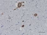
IHC staining of purified anti-α-Synuclein, 115-121 (clone LB... 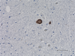
IHC staining of purified anti-α-Synuclein, 115-121 (clone LB... 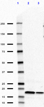
Western blot of purified anti-α-Synuclein, 115-121 antibody ... -
Biotin anti-α-Synuclein, 115-121
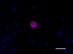
IHC staining of Biotin anti-anti-α-Synuclein, 115-121 antibo... 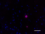
IHC staining of Biotin anti-anti-α-Synuclein, 115-121 antibo...
 Login / Register


Follow Us