- Clone
- 24G3 (See other available formats)
- Regulatory Status
- RUO
- Other Names
- PlGF, PLGF, Placental Growth Factor
- Isotype
- Mouse IgG2a
- Ave. Rating
- Submit a Review
| Cat # | Size | Price | Quantity Check Availability | Save | ||
|---|---|---|---|---|---|---|
| 534904 | 100 µg | 265 CHF | ||||
Placental growth factor (PlGF) is a member of the PDGF/VEGF family of growth factors. Four alternatively-spliced isoforms have been identified: PlGF-1, PlGF-2, PlGF-3, and PlGF-4. All isoforms are expressed in placental tissues, with PlGF-2 being expressed specifically during early placental development. PlGF-1 and PlGF-3 are considered diffusible isoforms, while PlGF-2 and PlGF-4 each contain a unique heparin binding domain that may increase associations with cell membranes. In addition to placental tissues, PlGF is expressed in the microvasculature, bone marrow, umbilical vein endothelia, uterine keratinocytes, and NK cells. PlGF binds to both VEGF-R1 and neuropilin, but not VEGF-R2 and induces monocyte activation, migration, inflammatory cytokine production. PlGF plays a key role in maternal vascular function during pregnancy, with circulating levels reaching a peak mid-gestation. It can be used as a marker for pregnancy-associated hypertensive disorders such as pre-eclampsia.
Product DetailsProduct Details
- Verified Reactivity
- Human
- Antibody Type
- Monoclonal
- Host Species
- Mouse
- Immunogen
- Recombinant protein
- Formulation
- Phosphate-buffered solution, pH 7.2, containing 0.09% sodium azide.
- Preparation
- The antibody was purified by affinity chromatography and conjugated with biotin under optimal conditions.
- Concentration
- 0.5 mg/ml
- Storage & Handling
- The antibody solution should be stored undiluted between 2°C and 8°C. Do not freeze.
- Application
-
ELISA-D - Quality tested
- Recommended Usage
-
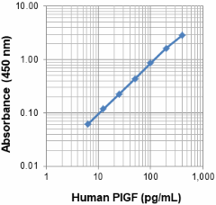
Each lot of this antibody is quality control tested by ELISA assay. For ELISA Detection applications, a concentration range of 0.1 - 4.0 µg/mL is recommended. It is recommended that the reagent be titrated for optimal performance for each application.
- Application Notes
-
The biotin 24G3 antibody is useful as the detection antibody in a sandwich ELISA assay, when used in conjunction with pure 13C8 antibody (Cat. No. 534802) as the capture antibody.
- RRID
-
AB_2894609 (BioLegend Cat. No. 534904)
Antigen Details
- Structure
- Homodimer
- Distribution
-
Vascular endothelial cells, smooth muscle cells, keratinocytes, hematopoitic cells, retinal pigment epithelial cells, tumor cells, and cerebellar stroma cells.
- Function
- PlGF is an angiogenic factor and induces proliferation and migration of endothelial cells. It stimulates endothelial cells and synergistically amplifies the action of VEGF. Soluble VEGFR1 (sFLT1) can regulate the accessibility of PlGF.
- Interaction
- Endothelial cells, Macrophages, and trophoblats.
- Ligand/Receptor
- VEGFR1 (FLT-1), and neuropilin 1 (Nrp1)
- Cell Type
- Endothelial cells, Epithelial cells, Hematopoietic stem and progenitors
- Antigen References
-
- Maglione D, et al. 1991. Proc Natl Acad Sci. USA 88:9267.
- Carmeliet P, et al. 2001. Nat Med. 7:575.
- Casalou C, et al. 2007. Leukemia. 21:1590.
- Marcellini M, et al. 2006. Am J Pathol. 169:643.
- Yao J, et al. 2011. Proc Natl Acad Sci USA. 108:11590.
- Cai J, et al. 2011. PLoS One. 6:e18076.
- Sibiude J, et al. 2012. PLoS One. 7:e50208.
- Snuderl M, et al. 2013. Cell. 152:1065.
- Gene ID
- 5228 View all products for this Gene ID
- UniProt
- View information about PlGF on UniProt.org
Related Pages & Pathways
Pages
Related FAQs
- How many biotin molecules are per antibody structure?
- We don't routinely measure the number of biotins with our antibody products but the number of biotin molecules range from 3-6 molecules per antibody.
Other Formats
View All PlGF Reagents Request Custom Conjugation| Description | Clone | Applications |
|---|---|---|
| Biotin anti-human PlGF | 24G3 | ELISA Detection |
Compare Data Across All Formats
This data display is provided for general comparisons between formats.
Your actual data may vary due to variations in samples, target cells, instruments and their settings, staining conditions, and other factors.
If you need assistance with selecting the best format contact our expert technical support team.


 Login / Register


Follow Us