CytoPhase™ Violet

Pricing & Availability
Regulatory Status
RUO
Other Names
live cell cycle
Ave. Rating
Submit a Review
Product Citations
publications
CytoPhase_Violet_103116
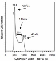
Ramos cells treated with 5µM CytoPhase™ Violet dye for 90 minutes at 37°C. Cells were then acquired on a flow cytometer equipped with a 405 nm laser with a 450/50 bandpass filter.
  • CytoPhase_Violet_103116
    Ramos cells treated with 5µM CytoPhase™ Violet dye for 90 minutes at 37°C. Cells were then acquired on a flow cytometer equipped with a 405 nm laser with a 450/50 bandpass filter.
See Cytophase™ Violet spectral data
Cat # Size Price Quantity Check Availability Save
425701 200 µL 282 CHF
Check Availability


Need larger quantities of this item?
Request Bulk Quote
Description

CytoPhase™ Violet is a cell-permeant DNA-binding dye with the molecular weight of 560.96D and can be excited by either UV (355 nm or 375 nm) or violet laser (405 nm). Its emission maximum wavelength is about 440nm. It can be used for flow cytometric analysis of cell cycle in live or fixed cells. This dye can also be used for counterstaining the nuclei of cells in fluorescent microscopic imaging.

Product Details
Technical Data Sheet (pdf)

Product Details

Formulation
5mM solution in H20 (200 µl/vial)
Concentration
5.0 mM
Storage & Handling
Upon receipt, CytoPhase™ Violet should be stored at 4°C and protected from light.
Application

FC - Quality tested

Recommended Usage

The recommended usage concentration for CytoPhase™ Violet is 5µM - 10µM.

Application Notes

Prepare cells of interest in appropriate cell culture media at a concentration of 0.5-1x 10⁶ cells /ml. Add CytoPhase™ Violet dye to the cell suspension to bring it to 5µM (1:1000)-10µM (1:500) working solution and incubate for 90 minutes at 37°C (CO2 incubator). Optimal concentration might require titration of the dye based on differing cell types. Transfer cells to a FACS tube (cells do not require a wash step) and acquire samples on a flow cytometer equipped with a violet laser.

CytoPhase™ Violet can be used concurrently with antibody staining procedures. 

Antigen Details

Distribution

Nucleus, DNA.

Biology Area
Cell Biology, Cell Cycle/DNA Replication, Cell Proliferation and Viability
Gene ID
NA

Related FAQs

There are no FAQs for this product.
Go To Top Version: 2    Revision Date: 05.20.2020

For Research Use Only. Not for diagnostic or therapeutic use.


This product is supplied subject to the terms and conditions, including the limited license, located at www.biolegend.com/terms) ("Terms") and may be used only as provided in the Terms. Without limiting the foregoing, BioLegend products may not be used for any Commercial Purpose as defined in the Terms, resold in any form, used in manufacturing, or reverse engineered, sequenced, or otherwise studied or used to learn its design or composition without express written approval of BioLegend. Regardless of the information given in this document, user is solely responsible for determining any license requirements necessary for user’s intended use and assumes all risk and liability arising from use of the product. BioLegend is not responsible for patent infringement or any other risks or liabilities whatsoever resulting from the use of its products.


BioLegend, the BioLegend logo, and all other trademarks are property of BioLegend, Inc. or their respective owners, and all rights are reserved.


8999 BioLegend Way, San Diego, CA 92121 www.biolegend.com
Toll-Free Phone: 1-877-Bio-Legend (246-5343) Phone: (858) 768-5800 Fax: (877) 455-9587

ProductsHere

Login / Register
Remember me
Forgot your password? Reset password?
Create an Account