 Login / Register


- Regulatory Status
- RUO
- Other Names
- Mitochondrial labeling
- Ave. Rating
- Submit a Review
- Product Citations
- publications

-

NIH3T3 cells were stained with 100 nM of MitoSpy™ Red CMXRos (red) for 20 minutes at 37°C, fixed with 1% paraformaldehyde (PFA) for ten minutes at room temperature, and permeabilized with 1X True Nuclear™ Perm Buffer for ten minutes at room temperature. Then the cells were stained with Flash Phalloidin™ NIR 647 (green) for 20 minutes at room temperature and counterstained with DAPI (blue). The image was captured with a 60x objective. -

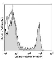
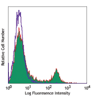
Human T-cell leukemia cell line, Jurkat, was treated with 1.0 µg/mL of LEAF™ purified anti-human CD95. At the end of incubation, cells were washed with PBS twice and stained with 25 nM of MitoSpy™ Red CMXRos for 20 minutes at 37°C. After incubation, cells were washed with Annexin V binding buffer twice and stained with 1.0 µg/T of Alexa Fluor® 488 Annexin V for ten minutes at room temperature.
| Cat # | Size | Price | Quantity Check Availability | Save | ||
|---|---|---|---|---|---|---|
| 424801 | 5 x 50 µg | 77 CHF | ||||
| 424802 | 20 x 50 µg | 247 CHF | ||||
MitoSpy™ mitochondrial localization probes are cell-permeant, fluorogenic chemical reagents that are used for labeling mitochondria in living cells. MitoSpy™ Red CMXRos localizes to the mitochondria based on its membrane potential and is useful to indicate cell health as well as for localization. It also can be fixed and permeabilized for further antibody-based detection.
Product DetailsProduct Details
- Verified Reactivity
- Human, Mouse, Rat, All Species
- Molecular Mass
- 531.52 g/mol
- Preparation
- The stock solution for MitoSpy™ Red CMXRos is prepared by dissolving the lyophilized probe in dimethyl sulfoxide (DMSO) to make a final concentration of 1mM by adding 94µl of DMSO to each vial.
- Storage & Handling
- Store MitoSpy™ Red CMXRos at -20°C.
- Application
-
ICC - Quality tested
FC - Verified - Recommended Usage
-
Each lot of this reagent is quality control tested by immunocytochemistry staining. For immunocytochemistry microscopy, a concentration range of 50 nM to 500 nM is recommended. For flow cytometric staining, a concentration range of 10 nM to 50 nM is recommended. It is recommended that the reagent be titrated for optimal performance for each application.
- Application Notes
-
MitoSpy™ Red CMXRos is excited at 577nm and emits at 598nm.
1. Prior to reconstitution, spin down the vial of lyophilized reagent in a microcentrofuge to ensure the reagent is at the bottom of the vial.
2. Reconstitute MitoSpy™ Red CMXRos to a 1mM concentration with DMSO by adding 94µl DMSO to an individual vial of lyophilized material. Protect the stock solution from light and keep frozen for storage.
3. Prepare the working solution for MitoSpy™ Red CMXRos in 37°C culture medium (incomplete), this will vary by cell line and type of imaging required.- If labeling mitochondria for live cell imaging a concentration between 50-250 nM is recommended.
- If cells are labeled live and then subsequently fixed a concentration between 250-500nM recommended.
4. Grow cells to a desired confluency and wash once with warm 1X PBS.
5. Add the diluted MitoSpy™ Red CMXRos solution to the live cells and place them in a 37°C incubator for 20-30 minutes.
6. Wash the cells twice with warm 1X PBS or culture media.
7. If the cells will be imaged live, they can now be imaged with a fluorescence microscope.
If the cells need to be fixed:
1. Fix the cells with 2-4% paraformaldehyde (PFA) for 10 mins at room temperature.
2. Wash the cells twice with 1X PBS.
3. Regular IF staining protocol can be used for antibodies or other probe co-stains.
If the cells need to be permeabilized:
1. Dilute the 10X True Nuclear™ 10X Perm Buffer in DI water.
2. Incubate cells with 1X perm buffer for 10 minutes at room temperature.
3. Wash cells with 1X PBS twice.
Antigen Details
- Distribution
-
Mitochondria.
- Biology Area
- Apoptosis/Tumor Suppressors/Cell Death, Cell Biology, Mitochondrial Function, Neuroscience
- Molecular Family
- Mitochondrial Markers
- Gene ID
- NA


Follow Us