- Clone
- NFLD.D2 (See other available formats)
- Regulatory Status
- RUO
- Other Names
- SS1, DRB1, DRw10, HLA-DRB, HLA-DR1B
- Isotype
- Mouse IgG1, κ
- Ave. Rating
- Submit a Review
- Product Citations
- publications
-

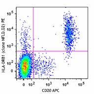
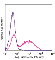
Human peripheral blood lymphoyctes were stained with CD20 APC and HLA-DRB1 (clone NFLD.D2, top) PE or mouse IgG1, κ PE isotype control (bottom). -

| Cat # | Size | Price | Quantity Check Availability | Save | ||
|---|---|---|---|---|---|---|
| 362303 | 25 tests | 165 CHF | ||||
| 362304 | 100 tests | 384 CHF | ||||
HLA-DRB1, a HLA class II beta chain paralog, is a heterodimer membrane anchored α (DRA) and β chain (DRB). It plays an imporant role in extracellular protein-derived peptide presentation. It is expressed by antigen presenting cells (APC) such as B cells, dendritic cells, and macrophages. The β chain (composed of β1 and β2) is 26-28 kD and contains polymorphisms allowing for variability in peptide binding specificities. DRB1 is expressed by all individuals and its expression is five fold higher than DRB chains 3, 4, or 5. The peptide binding groove can accommodate peptides 10-30 residues long. Thirty-three different sub-types of HLA-DRB1*04 have been described. Of these, HLA-DRB1*04 is associated with an increased incidence of rheumatoid arthritis and is present on a subset of DRB1 molecules.
Product DetailsProduct Details
- Verified Reactivity
- Human
- Antibody Type
- Monoclonal
- Host Species
- Mouse
- Immunogen
- L-cell transfectants HLA-DRB1*0401(11th IHW 8115), CFA
- Formulation
- Phosphate-buffered solution, pH 7.2, containing 0.09% sodium azide and BSA (origin USA)
- Preparation
- The antibody was purified by affinity chromatography and conjugated with PE under optimal conditions.
- Concentration
- Lot-specific (to obtain lot-specific concentration and expiration, please enter the lot number in our Certificate of Analysis online tool.)
- Storage & Handling
- The antibody solution should be stored undiluted between 2°C and 8°C, and protected from prolonged exposure to light. Do not freeze.
- Application
-
FC - Quality tested
- Recommended Usage
-
Each lot of this antibody is quality control tested by immunofluorescent staining with flow cytometric analysis. For flow cytometric staining, the suggested use of this reagent is 5 µl per million cells in 100 µl staining volume or 5 µl per 100 µl of whole blood.
- Excitation Laser
-
Blue Laser (488 nm)
Green Laser (532 nm)/Yellow-Green Laser (561 nm)
- Application Notes
-
Clone NFLD.D2 recognizes a shared epitope (DRB1*70-74:QKRAA and QRRAA) of HLA_DRB1*04.
-
Application References
(PubMed link indicates BioLegend citation) -
- Wicks I, et al. 1997. Ann. Rheum Dis. 56:135 (FC)
- Patil NS, et al. 2001. J. Immunol. 12:7157 (FC)
- Drover S, et al. 1998. Hum. Immunol. 59:77 (FA)
- RRID
-
AB_2563518 (BioLegend Cat. No. 362303)
AB_2563518 (BioLegend Cat. No. 362304)
Antigen Details
- Structure
- MHC class II cell surface receptor, 30 kD
- Distribution
- APCs
- Function
- Presentation of the peptide to T cells leads to activation or suppression of the immune system, which eventually leads to the immune response.
- Interaction
- CD4+ T cells
- Cell Type
- Antigen-presenting cells, B cells, Dendritic cells
- Biology Area
- Immunology, Innate Immunity
- Molecular Family
- MHC Antigens
- Antigen References
-
1. Scally SW, et al. 2013. J. Exp. Med. 210:2569.
2. Balnyte R, et al. 2013. BMC Neurol. 13:77.
3. Ranasinghe S, et al. 2013. Nat. Med. 19:930.
4. Foo JN, et al. 2013. Am. J. Hum. Genet. 93:167.
5. Balandraud N, et al. 2013. PLoS One. 8:e64108.
6. Sutton VR, et al. 1988. Hum. Immunol. 22:123.
7. Stunz LL, et al. 1989. J. Immunol. 143:3081. - Gene ID
- 3123 View all products for this Gene ID
- UniProt
- View information about HLA-DRB1 on UniProt.org
Related Pages & Pathways
Pages
Related FAQs
- What type of PE do you use in your conjugates?
- We use R-PE in our conjugates.
Other Formats
View All HLA-DRB1 Reagents Request Custom Conjugation| Description | Clone | Applications |
|---|---|---|
| Purified anti-human HLA-DRB1 | NFLD.D2 | FC |
| PE anti-human HLA-DRB1 | NFLD.D2 | FC |
Customers Also Purchased
Compare Data Across All Formats
This data display is provided for general comparisons between formats.
Your actual data may vary due to variations in samples, target cells, instruments and their settings, staining conditions, and other factors.
If you need assistance with selecting the best format contact our expert technical support team.
-
Purified anti-human HLA-DRB1
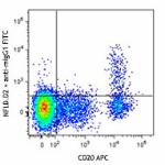
Human peripheral blood lymphoyctes were stained with CD20 AP... 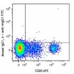
-
PE anti-human HLA-DRB1
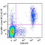
Human peripheral blood lymphoyctes were stained with CD20 AP... 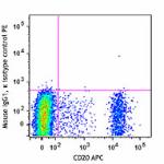
 Login / Register




Follow Us