- Clone
- W17035A (See other available formats)
- Regulatory Status
- RUO
- Other Names
- Cyclic AMP-dependent transcription factor ATF-6 beta, ATF-6 beta, Activating transcription factor 6 beta, ATF6-beta
- Isotype
- Rat IgG2b, κ
- Ave. Rating
- Submit a Review
- Product Citations
- publications

-

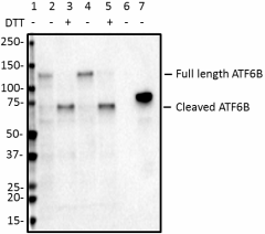
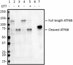
Western blot of anti-ATF6β antibody (clone W17035A). Lane 1: Molecular weight marker; Lane 2: 20 µg of Hela cell lysate; Lane 3: 20 µg lysate from Hela cells treated with 4mM DTT for 2 hours; Lane 4: 20 µg of SH-SY5Y cell lysate; Lane 5: 20 µg of lysate from SH-SY5Y cells treated with 4 mM DTT for 2 hours; Lane 6: 1 ng of His-tagged human ATF6α; Lane 7: 1 ng of His-tagged human ATF6β. The blot was incubated with 1 µg/mL of the primary antibody overnight at 4°C, followed by incubation with HRP-labeled goat anti-Rat IgG (Cat. No. 405405). Enhanced chemiluminescence was used as the detection system. -

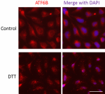
ICC staining of anti-ATF6β antibody (clone W17035A) on Hela cells. The cells were treated with (bottom) or without (top) 4 mM DTT for 2 hours, fixed with 4% PFA, permeabilized with a buffer containing 0.1% Triton X-100 and 0.25% BSA, and blocked with 2% normal goat serum and 0.02% BSA. The cells were then incubated with 5 µg/ml of the primary antibody overnight at 4°C, followed by incubation wit 5 µg/ml of Alexa Fluor® 594 goat anti-Rat IgG (Cat. No. 405422) for one hour at room temperature. Nuclei were counterstained with DAPI. The images were captured with a 40X objective. Scale bar: 50 µm
| Cat # | Size | Price | Quantity Check Availability | Save | ||
|---|---|---|---|---|---|---|
| 853201 | 25 µg | 112 CHF | ||||
| 853202 | 100 µg | 276 CHF | ||||
ATF6 encodes a transcription factor that is anchored in the endoplasmic reticulum (ER) and activated during the unfolded protein response (UPR) to protect cells from ER stress. Under conditions of ER stress, ATF6 is transported from the ER to the Golgi apparatus, where it is cleaved by Golgi-resident proteases to release the cytosolic DNA-binding portion. Then the processed ATF6 moves to the nucleus to activate gene expression. Deletion of the isoform activating transcription factor 6α (ATF6α) and its paralog ATF6β results in embryonic lethality and notochord dysgenesis in nonhuman vertebrates, and loss-of-function mutations in ATF6α are associated with malformed neuroretina and congenital vision loss in humans. Altered ATF6 function in neurodegenerative disorders such as Parkinson's disease has been observed.
Product DetailsProduct Details
- Verified Reactivity
- Human
- Antibody Type
- Monoclonal
- Host Species
- Rat
- Immunogen
- Human ATF6β recombinant protein (1-250 a.a.) expressed in E. coli.
- Formulation
- Phosphate-buffered solution, pH 7.2, containing 0.09% sodium azide.
- Preparation
- The antibody was purified by affinity chromatography.
- Concentration
- 0.5 mg/ml
- Storage & Handling
- The antibody solution should be stored undiluted between 2°C and 8°C.
- Application
-
WB - Quality tested
ICC - Verified - Recommended Usage
-
Each lot of this antibody is quality control tested by Western blotting. For Western blotting, the suggested use of this reagent is 1.0 - 5.0 µg per ml. For immunocytochemistry, a concentration range of 5.0 - 10.0 μg/ml is recommended. It is recommended that the reagent be titrated for optimal performance for each application.
- RRID
-
AB_2728607 (BioLegend Cat. No. 853201)
AB_2728608 (BioLegend Cat. No. 853202)
Antigen Details
- Structure
- ATF6β is a 703 amino acid protein with a molecular weight mass of ~80 kD.
- Distribution
-
Tissue distribution: ATF6β is ubiquitously expressed in numerous cell types including cells in of the central nervous system (CNS).
Cellular distribution: Cytoplasmic, nucleus, ER, and golgi apparatus.
- Function
- ATF6β is a transmembrane glycoprotein that functions as a transcription activator and initiates the unfolded protein response during ER stress.
- Interaction
- ATF6β interacts with ER chaperone immunoglobulin-binding protein (BiP), and can be cleaved by lumenal site 1 protease (S1P) and intra-membrane site 2 protease (S2P).
- Biology Area
- Cell Biology, Neurodegeneration, Neuroscience, Neuroscience Cell Markers, Protein Trafficking and Clearance, Transcription Factors
- Molecular Family
- Endoplasmic Reticulum Markers
- Antigen References
-
- Shen J, et al. 2002. Dev Cell. 3:99-111
- Ron D and Walter P. 2007. Nat Rev Mol Cell Biol. 8: 519-529
- Credle JJ, et al. 2015. Neurobiol Dis. 76: 112-125
- Jin JK, et al. 2017. Circ Res. 120: 862-875
- Kroeger H, et al. 2018. Sci Signal. 11: 5785
- Gene ID
- 1388 View all products for this Gene ID
- UniProt
- View information about ATF6beta on UniProt.org
Related Pages & Pathways
Pages
Related FAQs
Other Formats
View All ATF6β Reagents Request Custom Conjugation| Description | Clone | Applications |
|---|---|---|
| Purified anti-ATF6β | W17035A | WB,ICC |
Compare Data Across All Formats
This data display is provided for general comparisons between formats.
Your actual data may vary due to variations in samples, target cells, instruments and their settings, staining conditions, and other factors.
If you need assistance with selecting the best format contact our expert technical support team.
 Login / Register


Follow Us