 Login / Register


- Clone
- 3A1 (See other available formats)
- Regulatory Status
- RUO
- Other Names
- AAA, ABETA, ABPP, AD1, CTFgamma, CVAP, PN-II, PN2, Amyloid beta A4 protein, preA4, protease nexin-II, peptidase nexin-II, beta-amyloid peptide, alzheimer disease amyloid protein, cerebral vascular amyloid peptide, APP, Amyloid Precursor Protein
- Isotype
- Mouse IgG1, κ
- Ave. Rating
- Submit a Review
- Product Citations
- publications

-

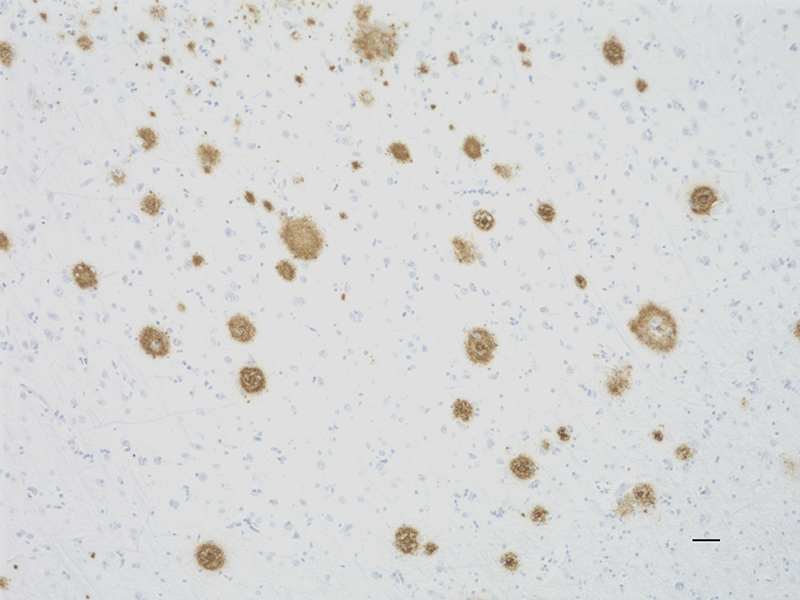
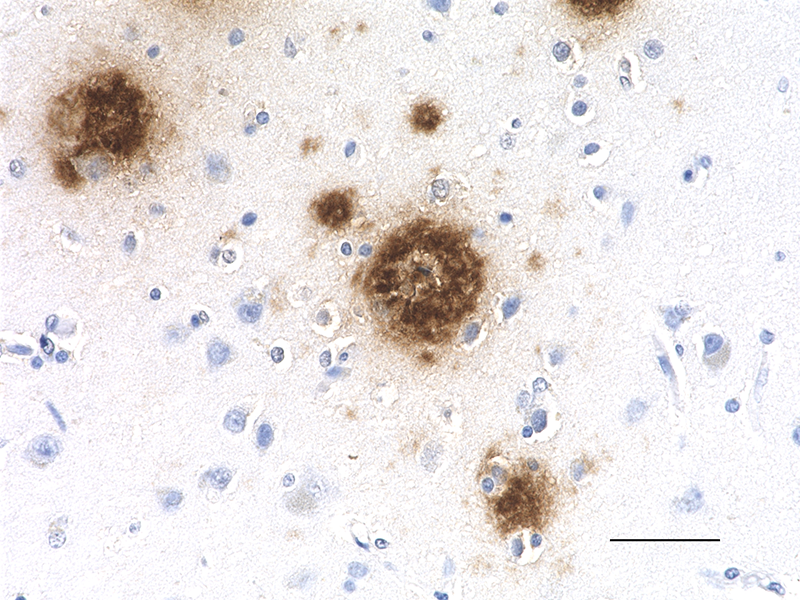
IHC staining of Purified anti-β-Amyloid, 1-15 (clone 3A1) on formalin-fixed paraffin-embedded Alzheimer’s disease human brain tissue. Following antigen retrieval using 88% formic acid, the tissue was incubated with primary antibody overnight at 4°C. BioLegend’s Ultra Streptavidin HRP Kit (Multi-Species, DAB, Cat. No. 929501) was used for detection followed by hematoxylin counterstaining. Images were captured with a 40X objective. Scale bar: 50 μm -

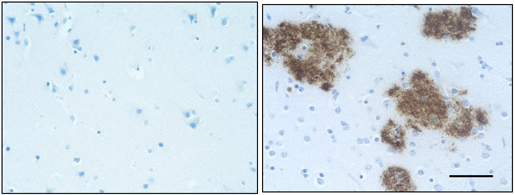
IHC staining of purified anti-β-Amyloid, 1-15 antibody (clone 3A1) on frozen normal (left panel) and Alzheimer's disease (right panel) human brain tissues. The tissues were incubated with 1 µg/mL of the primary antibody overnight at 4°C. BioLegend’s Ultra Streptavidin HRP Detection Kit (Cat. No. 929901) was used for detection followed by hematoxylin and bluing solution counterstaining, according to the protocol provided. The images were captured with a 40X objective. Scale bar: 50 µm. -

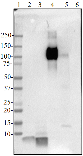
Direct ELISA of purified anti-β-Amyloid, 1-15 antibody (clone 3A1) binding to plate-immobilized Aβ1-40 (left panel) and lack of reactivity with plate-immobilized recombinant APP (right panel). ELISA was performed by coating wells with 100 ng of peptide or protein. The wells were then incubated with clones 3A1, 6E10 or mouse IgG at 37°C for 45 minutes, followed by incubation with horseradish peroxidase labeled goat anti-mouse secondary antibody. Clone 6E10 was included as a control for cross-reactivity with Aβ1-40 and APP. TMB (3, 3', 5, 5' tetramethylbenzidine, Cat. No. 421501) was used as the detection system.
| Cat # | Size | Price | Quantity Check Availability | Save | ||
|---|---|---|---|---|---|---|
| 847501 | 25 µg | 129 CHF | ||||
| 847502 | 100 µg | 323 CHF | ||||
Alzheimer’s disease is characterized by the accumulation of aggregated Aβ peptides in senile plaques and vascular deposits. Aβ peptides are derived from amyloid precursor protein (APP) through sequential proteolytic cleavage of APP by β- and γ-secretases generating diverse Aβ species. Aβ can aggregate to form soluble oligomeric species and insoluble fibrillar or amorphous assemblies. Some forms of the aggregated peptides are toxic to neurons.
Product DetailsProduct Details
- Verified Reactivity
- Human
- Antibody Type
- Monoclonal
- Host Species
- Mouse
- Immunogen
- This antibody was raised against dityrosine cross-linked human Aβ1-40.
- Formulation
- Phosphate-buffered solution, pH 7.2, containing 0.09% sodium azide.
- Preparation
- The antibody was purified by affinity chromatography.
- Concentration
- 0.5 mg/mL
- Storage & Handling
- The antibody solution should be stored undiluted between 2°C and 8°C.
- Application
-
IHC-P - Quality tested
IHC-F, Direct ELISA - Verified - Recommended Usage
-
Each lot of this antibody is quality control tested by formalin-fixed paraffin-embedded immunohistochemical staining. For immunohistochemistry on formalin-fixed paraffin-embedded tissue sections, a concentration range of 1.0 - 5.0 µg/mL is suggested. For immunohistochemistry on frozen tissue sections, a concentration range of 1.0 - 5.0 µg/mL is suggested. For ELISA applications, a concentration range of 0.01 - 1.0 µg/mL is recommended. It is recommended that the reagent be titrated for optimal performance for each application.
-
Application References
(PubMed link indicates BioLegend citation) -
- Frost JL, et al. 2015. Neurobiol Aging. 36(12):3187 (IHC-P)
- Lardenoije R, et al. 2018. Mol. Cell. Neurosci. 86:1-15. (IHC-P) PubMed
- RRID
-
AB_2728567 (BioLegend Cat. No. 847501)
AB_2728568 (BioLegend Cat. No. 847502)
Antigen Details
- Structure
- Aβ denotes peptides of 36-43 amino acids generated from cleavage of APP by secretases. Aβ has an apparent molecular mass of about 4 kD.
- Distribution
-
Tissue distribution: Primarily nervous system, but also adipose tissue, intestine, muscle.
Cellular distribution: Cytosol, endosomes, nucleus, plasma membrane, extracellular, and golgi apparatus.
- Function
- The normal function of Aβ is not well understood. Several potential physiological roles have been proposed, including: activation of kinase enzymes; protection against oxidative stress; regulation of cholesterol transport; transcription factor, and as an anti-microbial agent.
- Interaction
- Tau, Prion
- Biology Area
- Cell Biology, Neurodegeneration, Neuroscience, Protein Misfolding and Aggregation
- Molecular Family
- APP/β-Amyloid
- Antigen References
-
- Puig KL, et al. 2012. Exp Gerontol. 48(7): 608.
- Selkoe DJ, et al. 2016. EMBO Mol Med. 8(6):595.
- Walsh DM, et a. 2007. J Neurochem. 101(5):1172.
- Gene ID
- 351 View all products for this Gene ID
- UniProt
- View information about beta-Amyloid on UniProt.org

Related FAQs
Other Formats
View All β-Amyloid Reagents Request Custom Conjugation| Description | Clone | Applications |
|---|---|---|
| Purified anti-β-Amyloid, 1-15 | 3A1 | IHC-P,IHC-F,Direct ELISA |
Customers Also Purchased
Compare Data Across All Formats
This data display is provided for general comparisons between formats.
Your actual data may vary due to variations in samples, target cells, instruments and their settings, staining conditions, and other factors.
If you need assistance with selecting the best format contact our expert technical support team.


Follow Us