- Clone
- W19011B (See other available formats)
- Regulatory Status
- RUO
- Other Names
- G0/G1 switch regulatory protein 3 (G0S3), FosB Proto-Oncogene, AP-1 Transcription Factor Subunit, Oncogene Fos-B, FBJ Murine Osteosarcoma Viral Oncogene Homolog B
- Isotype
- Rat IgG2a, κ
- Ave. Rating
- Submit a Review
- Product Citations
- publications
-

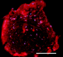
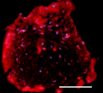
IHC staining of purified anti-FosB (clone W19011B) on formalin-fixed paraffin-embedded human thymus tissue. Following antigen retrieval using 1x Tris-Buffered Saline (final concentration 0.05M) w/ Tween 20 (Cat. No. 925501), the tissue was incubated with 5 μg/mL of the primary antibody overnight at 4°C, followed by incubation with 2.5 μg/mL of Alexa Fluor® 647 goat anti-rat IgG (Cat. No. 405416) (red) for one hour at room temperature. Nuclei were counterstained with DAPI (blue), and the slide was mounted with ProLong™ Gold Antifade Mountant. The image was captured with a 20x objective. Scale bar: 50 μm -

ICC staining of purified anti-FosB (clone W19011B) (panel A) or purified rat IgG2a, κ isotype control (Cat. No. 400501) (panel B) on HeLa cells treated with PMA (100 ng/mL) for 4 hours. The cells were fixed with 4% PFA, permeabilized with 0.5% Triton-X, and blocked with 5% FBS for 1 hour at room temperature. The cells were then stained with 5.0 μg/mL of the primary antibody, followed by incubation with 2.5 μg/mL of Alexa Fluor® 594 goat anti-rat IgG (Cat. No. 405422) for 1 hour at room temperature. Nuclei were counterstained with DAPI, and the image was captured with a 40X objective. Scale bar = 50 μm -

Hela cells treated with 100 ng PMA for 4 hours (positive target) were fixed with Fixation Buffer (Cat. No. 420801), permeabilized using True-Phos™ Perm Buffer (Cat. No. 425401), and intracellularly stained with 0.50 μg of purified anti-FosB (clone W19011B) (filled histogram) or 0.50 μg of purified rat IgG2a, κ isotype control (Cat. No. 400502) (open histogram) followed by PE goat anti-rat IgG (Cat. No. 405406). -

Untreated HeLa cells (negative target) were fixed with Fixation Buffer (Cat. No. 420801), permeabilized using True-Phos™ Perm Buffer (Cat. No. 425401), and intracellularly stained with 0.50 μg of purified anti-FosB (clone W19011B) (filled histogram) or 0.50 μg of purified rat IgG2a, κ isotype control (Cat. No. 400501) (open histogram) followed by PE goat anti-rat IgG (Cat. No. 405406).
| Cat # | Size | Price | Quantity Check Availability | Save | ||
|---|---|---|---|---|---|---|
| 600851 | 25 µg | 118 CHF | ||||
| 600852 | 100 µg | 293 CHF | ||||
FosB is a member of the FOS family of proteins that dimerizes with proteins of the Jun family to form the AP-1 transcription factor complex. As part of AP-1, FosB plays a critical role in regulating cell differentiation, proliferation, and apoptosis. FosB is expressed in most tissues and has been particularly well-studied in the CNS. ΔFosB, a truncated splice variant of the FOSB gene, plays a critical role in the brain’s reward circuit plasticity, with significant impact on both natural and pathological behavior. FosB also functions as a tumor suppressor, and dysregulation of FosB activity or levels has been found in numerous cancers, including gastric, prostate and colorectal cancer.
Product DetailsProduct Details
- Verified Reactivity
- Human
- Antibody Type
- Monoclonal
- Host Species
- Rat
- Immunogen
- Full-length recombinant FosB protein
- Formulation
- Phosphate-buffered solution, pH 7.2, containing 0.09% sodium azide
- Preparation
- The antibody was purified by affinity chromatography.
- Concentration
- 0.5 mg/mL
- Storage & Handling
- The antibody solution should be stored undiluted between 2°C and 8°C.
- Application
-
IHC-P - Quality tested
ICC, ICFC - Verified - Recommended Usage
-
Each lot of this antibody is quality control tested by formalin-fixed paraffin-embedded immunohistochemical staining. For immunohistochemistry, a concentration range of 5 - 10 µg/mL is suggested. For immunocytochemistry, a concentration range of 5 - 10 μg/mL is recommended. For intracellular flow cytometric staining, the suggested use of this reagent is ≤ 0.25 µg per million cells in 100 µL volume. It is recommended that the reagent be titrated for optimal performance for each application.
- Application Notes
-
For ICC, we recommend fixation in 4% PFA and permeabilization for 6 minutes in 0.5% Triton-X.
Methanol only and PFA + Methanol fix/perm methods were tested and produced inferior signal. - RRID
-
AB_2922629 (BioLegend Cat. No. 600851)
AB_2922629 (BioLegend Cat. No. 600852)
Antigen Details
- Structure
- FosB is a 338 amino acid protein with a predicted molecular weight of 36kD.
- Distribution
-
FosB is widely expressed in developing and adult tissues, including the brain, thyroid, bladder and adipose tissue.
- Function
- FosB heterodimerizes with proteins of the Jun family to form the transcription factor AP-1 to regulate cell proliferation, survival, and differentiation.
- Interaction
- Jun family proteins e.g. c-Jun, genes with the TPA response element (TGA G/C TCA) SWI/SNF chromatin remodeling complex, DPP4
- Cell Type
- Mature Neurons
- Biology Area
- Cell Biology, Cell Proliferation and Viability, Neuroscience, Neuroscience Cell Markers, Transcription Factors
- Molecular Family
- Tumor Suppressors
- Antigen References
-
- Nestler E. 2015. Eur J Pharmacol. 753:66-72.
- Milde-Langosch K. 2005. Eur J Cancer. 41:2449-61.
- Gene ID
- 2354 View all products for this Gene ID
- UniProt
- View information about FosB on UniProt.org
Related Pages & Pathways
Pages
Related FAQs
Other Formats
View All FosB Reagents Request Custom Conjugation| Description | Clone | Applications |
|---|---|---|
| Purified anti-FosB | W19011B | IHC-P,ICC,ICFC |
| Alexa Fluor® 647 anti-FosB | W19011B | ICFC,ICC |
| PE anti-FosB | W19011B | ICFC |
Compare Data Across All Formats
This data display is provided for general comparisons between formats.
Your actual data may vary due to variations in samples, target cells, instruments and their settings, staining conditions, and other factors.
If you need assistance with selecting the best format contact our expert technical support team.
-
Purified anti-FosB
IHC staining of purified anti-FosB (clone W19011B) on formal... 
ICC staining of purified anti-FosB (clone W19011B) (panel A)... 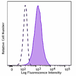
Hela cells treated with 100 ng PMA for 4 hours (positive tar... 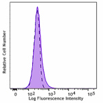
Untreated HeLa cells (negative target) were fixed with Fixat... -
Alexa Fluor® 647 anti-FosB
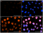
HeLa cells untreated (panels A and B) or treated with 100 ng... 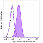
HeLa cells untreated (negative control, open histogram) or t... -
PE anti-FosB
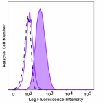
HeLa cells untreated (negative control, open histogram) or t...
 Login / Register


Follow Us