 Login / Register


- Clone
- A19063A (See other available formats)
- Regulatory Status
- RUO
- Other Names
- Leu-19, NKH1, NCAM-1
- Isotype
- Mouse IgG2b, κ
- Ave. Rating
- Submit a Review
- Product Citations
- publications

-

Human paraffin-embedded cerebellum tissue slices were prepared with a standard protocol of deparaffination and rehydration. Antigen retrieval was done with citrate buffer 1X pH6.0 at 95°C for 40 minutes. Tissue was washed with PBS/ 0.05% Tween-20 twice for five minutes, blocked with 5% FBS and 0.2% gelatin for 30 minutes. Then, the tissue was stained with 10 µg/mL of purified anti-human CD56 (NCAM) (clone A19063A) at 4°C overnight. On the next day, the tissue was washed twice with PBS and stained with Alexa Fluor® 594 Goat anti-mouse IgG (minimal x-reactivity) (clone Poly4053) (red) for two hours at room temperature. The nuclei were counterstained with DAPI (blue). The image was scanned with a 10X objective and stitched with software. -

Human paraffin-embedded colon tissue slices were prepared with a standard protocol of deparaffination and rehydration. Antigen retrieval was done with citrate buffer 1X pH6.0 at 95°C for 40 minutes. Tissue was washed with PBS/ 0.05% Tween-20 twice for five minutes, blocked with 5% FBS and 0.2% gelatin for 30 minutes. Then, the tissue was stained with 10 µg/mL of purified anti-human CD56 (NCAM) (clone A19063A) at 4°C overnight. On the next day, the tissue was washed twice with PBS and stained with Alexa Fluor® 594 Goat anti-mouse IgG (minimal x-reactivity) (clone Poly4053) (red) for two hours at room temperature. The nuclei were counterstained with DAPI (blue). The image was scanned with a 10X objective and stitched with software. -

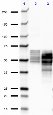
Whole cell extracts (15 µg total protein) from the indicated cell lines were resolved on a 4-12% Bis-Tris gel, transferred to a PVDF membrane, and probed with 1 µg/mL (1:500 dilution) of purified anti-human CD56 (NCAM) (clone A19063A) overnight at 4°C. Proteins were visualized by chemiluminescence detection using HRP goat anti-mouse IgG (Cat. No. 405306) at a 1:3000 dilution. Direct-Blot™ HRP anti-β-actin (Cat. No. 664804) was used as a loading control at a 1:25000 dilution (lower). Western-Ready™ ECL Substrate Plus Kit (Cat. No. 426317) was used as a detection agent. Lane M: Molecular weight marker -

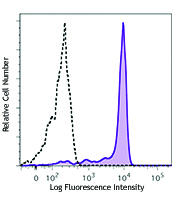
ICC staining of purified anti-human CD56 (NCAM) (clone A19063A) on HeLa cells. The cells were fixed and permeabilized with ice-cold methanol, and blocked with 5% FBS for 1 hour at room temperature. The cells were then stained with 1.0 µg/mL of the primary antibody, followed by incubation with 2.5 µg/mL of Alexa Fluor® 594 goat anti-mouse IgG (Cat. No. 405326) for 1 hour at room temperature. Nuclei were counterstained with DAPI (Cat. No. 422801), and the image was captured with a 40X objective. -

ICC staining of mouse IgG2b, κ isotype control (Cat. No. 400301) on HeLa cells. The cells were fixed and permeabilized with ice-cold methanol, and blocked with 5% FBS for 1 hour at room temperature. The cells were then stained with 1.0 µg/mL of the primary antibody, followed by incubation with 2.5 µg/mL of Alexa Fluor® 594 goat anti-mouse IgG (Cat. No. 405326) for 1 hour at room temperature. Nuclei were counterstained with DAPI (Cat. No. 422801), and the image was captured with a 40X objective.
| Cat # | Size | Price | Quantity Check Availability | Save | ||
|---|---|---|---|---|---|---|
| 380702 | 100 µg | 136 CHF | ||||
CD56 is a single transmembrane glycoprotein also known as NCAM (neural cell adhesion molecule), Leu-19, or NKH1. It is a member of the Ig superfamily. The 140 kD isoform is expressed on NK and NKT cells. CD56 is also expressed in the brain (cerebellum and cortex) and at neuromuscular junctions. Certain large granular lymphocyte (LGL) leukemias, small-cell lung carcinomas, neuronal derived tumors, myelomas, and myeloid leukemias also express CD56. CD56 plays a role in homophilic and heterophilic adhesion via binding to itself or heparan sulfate.
Product DetailsProduct Details
- Verified Reactivity
- Human
- Antibody Type
- Monoclonal
- Host Species
- Mouse
- Formulation
- Phosphate-buffered solution, pH 7.2, containing 0.09% sodium azide
- Preparation
- The antibody was purified by affinity chromatography.
- Concentration
- 0.5 mg/mL
- Storage & Handling
- The antibody solution should be stored undiluted between 2°C and 8°C.
- Application
-
IHC-P - Quality tested
ICC, WB - Verified - Recommended Usage
-
Each lot of this antibody is quality control tested by formalin-fixed paraffin-embedded immunohistochemical staining. For immunohistochemistry, a concentration range of 5.0 - 10.0 µg/mL is suggested. For immunocytochemistry, a concentration range of 0.1 - 10.0 μg/mL is recommended. For western blotting, the suggested use of this reagent is 1.0 - 5.0 µg/mL. It is recommended that the reagent be titrated for optimal performance for each application.
- RRID
-
AB_2922611 (BioLegend Cat. No. 380702)
Antigen Details
- Structure
- Ig superfamily, single transmembrane or GPI-anchored glycoprotein
- Distribution
-
NK cells, T subset, neural tissue, some LGL and myeloid leukemias
- Function
- Adhesion
- Ligand/Receptor
- Heparan sulfate
- Cell Type
- B cells, Leukemia, Mesenchymal Stem Cells, Neurons, NK cells, T cells
- Biology Area
- Cell Adhesion, Cell Biology, Costimulatory Molecules, Immunology, Innate Immunity, Neuroscience, Stem Cells, Synaptic Biology
- Molecular Family
- Adhesion Molecules, CD Molecules
- Antigen References
-
- Laroni A, et al. 2016. J Autoimmun. 72:8-18. PubMed
- Pachnio A, et al. 2016. PLoS Pathog. 12:e1005832. PubMed
- Wouters K, et al. 2017. Sci Rep. 7:42665. PubMed
- Gene ID
- 4684 View all products for this Gene ID
- UniProt
- View information about CD56 on UniProt.org


Related FAQs
Other Formats
View All CD56 Reagents Request Custom Conjugation| Description | Clone | Applications |
|---|---|---|
| Purified anti-human CD56 (NCAM) | A19063A | IHC-P,ICC,WB |
| Alexa Fluor® 647 anti-human CD56 (NCAM) | A19063A | IHC-P |
Customers Also Purchased
Compare Data Across All Formats
This data display is provided for general comparisons between formats.
Your actual data may vary due to variations in samples, target cells, instruments and their settings, staining conditions, and other factors.
If you need assistance with selecting the best format contact our expert technical support team.
-
Purified anti-human CD56 (NCAM)

Human paraffin-embedded cerebellum tissue slices were prepar... 
Human paraffin-embedded colon tissue slices were prepared wi... 
Whole cell extracts (15 µg total protein) from the indicated... 
ICC staining of purified anti-human CD56 (NCAM) (clone A1906... 
ICC staining of mouse IgG2b, κ isotype control (Cat. No. 400... -
Alexa Fluor® 647 anti-human CD56 (NCAM)

IHC staining of anti-human CD56 (clone A19063A) Alexa Fluor®... 
IHC staining of anti-human CD56 (clone A19063A) Alexa Fluor®...


Follow Us