- Clone
- W17101A (See other available formats)
- Regulatory Status
- RUO
- Other Names
- Receptor-Interacting Protein Kinase 1, RIP, RIP-1, RIP1, IMD57, AIEFL, Receptor-Interacting Serine/Threonine-Protein Kinase
- Isotype
- Rat IgG2b, λ
- Ave. Rating
- Submit a Review
- Product Citations
- publications

-

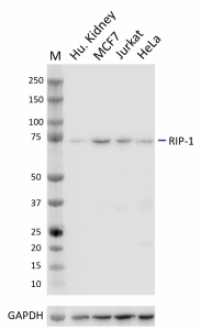
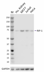
Whole cell extracts (15 µg total protein) from the indicated cell lines were resolved by 4-12% Bis-Tris gel electrophoresis, transferred to a PVDF membrane, and probed with 0.5 μg/mL (1:1000 dilution) purified anti-RIP-1 (clone W17101A) overnight at 4°C. Proteins were visualized by chemiluminescence detection using HRP goat anti-rat IgG (Cat. No. 405405) at a 1:3000 dilution. Western-Ready™ ECL Substrate Premium Kit (Cat. No. 426319) was used as a detection agent. Direct-Blot™ HRP anti-GAPDH (Cat. No. 607904) was used as a loading control at a 1:25000 dilution. Lane M: Molecular weight marker -

ICC staining of purified anti-RIP-1 (clone W17101A) on HeLa cells. The cells were fixed and permeabilized with ice-cold methanol and blocked with 5% FBS for 1 hour at room temperature. The cells were then stained with 5.0 µg/mL of the primary antibody, followed by incubation with 2.5 µg/mL of Alexa Fluor® 594 goat anti-rat IgG (Cat. No. 405422) for 1 hour at room temperature. Nuclei were counterstained with DAPI (Cat. No. 422801), and the image was captured with a 60X objective. Scale bar: 10 µm
| Cat # | Size | Price | Quantity Check Availability | Save | ||
|---|---|---|---|---|---|---|
| 603351 | 25 µg | 118 CHF | ||||
| 603352 | 100 µg | 293 CHF | ||||
Receptor-interacting serine/threonine-protein kinase 1 (RIP-1) is a member of the RIP family of serine/threonine kinases and consists of a kinase domain (KD) at its N-terminus, and a death domain (DD) in its C-terminus. It is a key regulator of cellular inflammatory response and cell survival when functioning as a scaffold protein, namely as part of the NF-κB signaling pathway, and is a key regulator of cell death when functioning as a kinase. RIP-1 promotes cell survival as part of the tumor necrosis factor receptor 1 (TNFR1) complex following ubiquitination by M1 and K63, and phosphorylation mediated by IKK. When ubiquitination is disrupted by ubiquitin E3 ligases, RIP-1 associates with FADD and caspase 8 to promote apoptosis or forms a complex with RIPK3 to induce necroptosis. Due to it being part of the TNF signaling pathway and its dual roles in cell survival and cell death, RIP-1 is of particular interest as a therapeutic target, namely as a mediator in the induction of immunogenic cell death and the treatment of cancer as well as in the treatment of autoimmune and neurodegenerative disorders.
Product DetailsProduct Details
- Verified Reactivity
- Human
- Antibody Type
- Monoclonal
- Host Species
- Rat
- Immunogen
- Partial recombinant human RIP1 protein
- Formulation
- Phosphate-buffered solution, pH 7.2, containing 0.09% sodium azide
- Preparation
- The antibody was purified by affinity chromatography.
- Concentration
- 0.5 mg/mL
- Storage & Handling
- The antibody solution should be stored undiluted between 2°C and 8°C.
- Application
-
WB - Quality tested
ICC - Verified - Recommended Usage
-
Each lot of this antibody is quality control tested by western blotting. For western blotting, the suggested use of this reagent is 0.5 µg/mL. For immunocytochemistry, a concentration of 5 μg/mL is recommended. It is recommended that the reagent be titrated for optimal performance for each application.
- Application Notes
-
This clone was tested for ICC using HeLa cells with localization expected in the cytosol and plasma membrane. Three fix/perm methods were used: methanol fixation, or 4% PFA fixation followed by permeabilization with either methanol or Triton X-100. Methanol-only fixation produced a strong signal with the expected cytoplasmic localization. PFA-fixation followed by permeabilization with either methanol or Triton X-100 did not produce the expected cytoplasmic/membrane signal.
This clone was tested for IHC-P on human skin tissue using Biolegend’s AEC Chromagen. Positive signal was not observed. - RRID
-
AB_2922634 (BioLegend Cat. No. 603351)
AB_2922634 (BioLegend Cat. No. 603352)
Antigen Details
- Structure
- RIP-1 is a 671 amino acid protein with a predicted molecular weight of 76 kD.
- Distribution
-
Ubiquitously expressed/localized to the cell membrane and cytoplasm
- Function
- Serine/threonine-protein kinase, Transferase
- Biology Area
- Apoptosis/Tumor Suppressors/Cell Death, Cell Biology, Cell Death, Neurodegeneration, Neuroscience
- Molecular Family
- Protein Kinases/Phosphatase
- Antigen References
-
- Ang RL, et al. 2019. Front Cell Dev Biol. 7:163.
- Degterev A, et al. 2019. Proc Natl Acad Sci U S A. 116:9714-22.
- Moriwaki K, et al. 2021. Mucosal Immunol. 15:84-95.
- Smith HG, et al. 2020. EMBO Mol Med. 12:e10979.
- Gene ID
- 8737 View all products for this Gene ID
- UniProt
- View information about RIP-1 on UniProt.org
Related FAQs
Other Formats
View All RIP-1 Reagents Request Custom Conjugation| Description | Clone | Applications |
|---|---|---|
| Purified anti-RIP-1 | W17101A | WB,ICC |
Compare Data Across All Formats
This data display is provided for general comparisons between formats.
Your actual data may vary due to variations in samples, target cells, instruments and their settings, staining conditions, and other factors.
If you need assistance with selecting the best format contact our expert technical support team.
 Login / Register


Follow Us