
 Login / Register


- Clone
- A17012A (See other available formats)
- Regulatory Status
- RUO
- Other Names
- Signal Transduced Activator of Transcription 1, Transcription Factor ISGF-3 components p91/p84
- Isotype
- Mouse IgG1, κ
- Ave. Rating
- Submit a Review


| Cat # | Size | Price | Quantity Check Availability | Save | ||
|---|---|---|---|---|---|---|
| 666401 | 25 µg | 112 CHF | ||||
| 666402 | 100 µg | 265 CHF | ||||
STAT1, also known as signal transduction and activator of transcription 1, is a ubiquitously expressed cytoplasmic protein and is activated in response to cytokine signaling, including IFN-α, IFN-γ, EGF, PDGF, and IL-6. Upon activation, STAT1 is phosphorylated at Tyrosine 701 (Tyr701) by receptor-associated kinases, including JAK1, JAK2, and TYK2. This results in STAT1 dimerization and subsequent translocation to the nucleus, where it functions as a transcriptional activator. STAT1 is involved in IFN-mediated immune responses, and STAT1-deficient mice are highly sensitive to bacterial and viral infections.
Product DetailsProduct Details
- Verified Reactivity
- Human
- Antibody Type
- Monoclonal
- Host Species
- Mouse
- Formulation
- Phosphate-buffered solution, pH 7.2, containing 0.09% sodium azide.
- Preparation
- The antibody was purified by affinity chromatography.
- Concentration
- 0.5 mg/ml
- Storage & Handling
- The antibody solution should be stored undiluted between 2°C and 8°C, and protected from prolonged exposure to light. Do not freeze.
- Application
-
WB - Quality tested
ICFC, ICC, ChIP - Verified - Recommended Usage
-
Each lot of this antibody is quality control tested by Western blotting. For Western blotting, the suggested use of this reagent is 0.1 - 2.0 µg per ml (1:250-5000 dilution). For intracellular flow cytometric staining, the suggested use of this reagent is ≤0.5 µg per million cells in 100 µl volume. For immunocytochemistry, a range of 5.0 μg/ml is recommended. For ChIP applications, the suggested dilution is 1:50 by volume. It is recommended that the reagent be titrated for optimal performance for each application.
For intracellular flow cytometry our True-Nuclear™ Transcription Factor Staining Protocol is recommended. - Application Notes
-
Clone A17012A recognizes STAT1 phosphorylated at Tyrosine 701 (Tyr701).
When using this clone for ICC, we recommend using methanol to permeabilize fixed cells. - RRID
-
AB_2728499 (BioLegend Cat. No. 666401)
AB_2728500 (BioLegend Cat. No. 666402)
Antigen Details
- Structure
- Two isoforms of STAT1 exist as a result of alternative splicing. Isoform α is a 750 amino acid protein with a predicted molecular weight of 87 kD; the β isoform is a 712 amino acid protein with a predicted molecular weight of 83 kD.
- Distribution
-
Ubiquitous tissue expression, nucleoplasmic-cytosolic distribution
- Function
- Immune response activation, cell signaling
- Interaction
- STAT1, STAT2, IRF9, ERBB4, JAK1, JAK2, TYK2, TNK1, SHP2
- Biology Area
- Cell Biology
- Molecular Family
- Nuclear Markers, Phospho-Proteins
- Antigen References
-
- Moretti S, et al. 2017. J. Biol. Chem. 292: 1785.
- Wei J, et al. 2015. J. Immunol. 195: 2870.
- Sung PS, et al. 2015. Proc. Natl. Acad. Sci. USA. 112: 10443
- Ooi EL, et al. 2014. Proc. Natl. Acad. Sci. USA. 111: 1909.
- Wu TR, et al. 2002. J. Biol. Chem. 277: 47572.
- Horvath, et al. 1996. J. Virol. 70: 647.
- Haque SJ, et al. 1995. J. Biol. Chem. 270: 25709.
- Gene ID
- 6772 View all products for this Gene ID
- UniProt
- View information about STAT1 Phospho Tyr701 on UniProt.org

Related FAQs
Other Formats
View All STAT1 Phospho (Tyr701) Reagents Request Custom Conjugation| Description | Clone | Applications |
|---|---|---|
| Purified anti-STAT1 Phospho (Tyr701) | A17012A | WB,ICFC,ICC,ChIP |
| PE anti-STAT1 Phospho (Tyr701) | A17012A | ICFC |
| Alexa Fluor® 647 anti-STAT1 Phospho (Tyr701) | A17012A | ICFC |
| PerCP/Cyanine5.5 anti-STAT1 Phospho (Tyr701) | A17012A | ICFC |
Customers Also Purchased
Compare Data Across All Formats
This data display is provided for general comparisons between formats.
Your actual data may vary due to variations in samples, target cells, instruments and their settings, staining conditions, and other factors.
If you need assistance with selecting the best format contact our expert technical support team.
-
Purified anti-STAT1 Phospho (Tyr701)

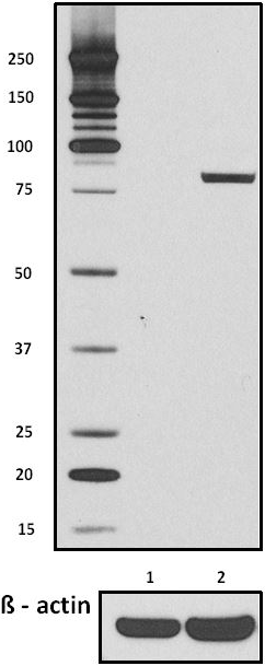
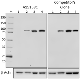
Total cell lysates (15 µg protein) from serum-starved HeLa S... 
Total cell lysates (15 µg protein) from serum-starved HeLa S... 
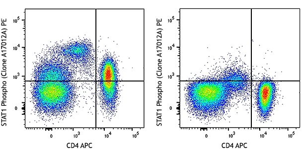
Human peripheral blood mononuclear cells were treated with (... 
Human peripheral blood monocytes were treated with (filled h... 
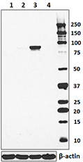
Serum starved HeLa cells were untreated (panel A) or stimula... 
Chromatin Immunoprecipitations (ChIP) were performed with cr... -
PE anti-STAT1 Phospho (Tyr701)

Human peripheral blood mononuclear cells were treated with (... 
Human peripheral blood monocytes were treated with (filled h... -
Alexa Fluor® 647 anti-STAT1 Phospho (Tyr701)

Human peripheral blood mononuclear cells were treated with (... 
Human peripheral blood monocytes were treated with (filled h... -
PerCP/Cyanine5.5 anti-STAT1 Phospho (Tyr701)

Human peripheral blood mononuclear cells were treated with (... 
Human peripheral blood monocytes were treated with (filled h...


Follow Us