
Discover the power of targeted proteomics using reagents you already trust

Bruker Spatial Biology presents CellScape™ Precise Spatial Multiplexing, an end-to-end immunofluorescence platform for highly multiplexed spatial proteomics and single-cell analysis. With advanced optical capabilities, streamlined fluidics for walk-away automation, and unprecedented flexibility in assay design, the CellScape system will accelerate your exploration into the rapidly evolving field of spatial biology.
CellScape technology features:
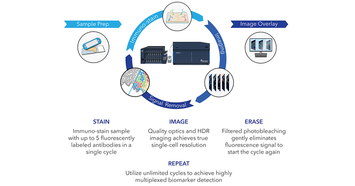
CellScape enables advanced quantitative analyses of every cell via built-in software and compatibility with third-party platforms for image processing and spatial analyses.
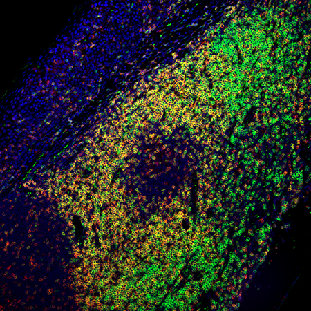
Multiplexed IHC staining of PerCP/Cyanine5.5 anti-CD45RO (clone UCHL1) on formalin-fixed paraffin-embedded human tonsil tissue, validated for use on the CellScape. The tissue was incubated with PerCP/Cyanine5.5 anti-CD45RO (clone UCHL1, red) and Alexa Fluor® 488 anti-CD3 (green) for one hour at room temperature. Nuclei were counterstained with Hoechst 33342. Images were captured with a 20X objective.
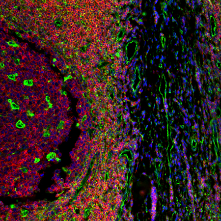
Multiplexed IHC staining of Alexa Fluor® 488 anti-Vimentin on formalin-fixed paraffin-embedded human tonsil tissue, validated for use on the Cellscape. The tissue was iteratively stained with Alexa Fluor® 488 anti-Vimentin (green) and Alexa Fluor® 488 anti-CD3 (red) for one hour at room temperature. Nuclei were counterstained with DAPI. Images were captured with a 20X objective.
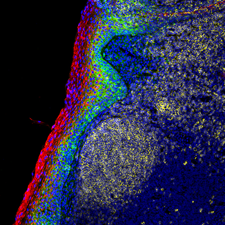
Multiplexed IHC staining of PerCP/Cyanine5.5 anti-CD138 (clone MI15) on formalin-fixed paraffin-embedded human tonsil tissue, validated for use on the Cellscape. The tissue was incubated with PerCP/Cyanine5.5 anti-CD138 (clone MI15, green) and PerCP/Cyanine5.5 anti-CD45 (red) for one hour at room temperature. Nuclei were counterstained with Hoechst 33342. Images were captured with a 20X objective.
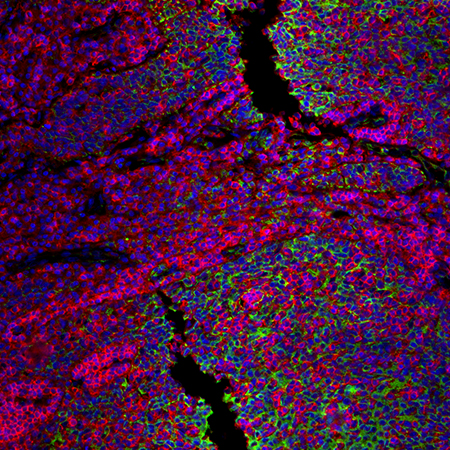
Multiplexed IHC staining of PerCP/Cyanine5.5 anti-CD326 on formalin-fixed paraffin-embedded human tonsil tissue, validated for use on the Cellscape. The tissue was iteratively stained with PerCP/Cyanine5.5 anti-CD326 (green) and PerCP/Cyanine5.5 anti-CD45 (red) for one hour at room temperature. Nuclei were counterstained with Hoechst 33342. Images were captured with a 20X objective.
CellScape can be utilized with pre-optimized panels, validated individual antibodies, or your own fluorescently labelled markers. In collaboration with Bruker Spatial Biology, Biolegend has produced and validated antibodies compatible with CellScape to expand the number of targets for your multiplexing research.
|
Specificity |
Clone |
|
NAT105 |
|
|
LG.3A10 |
|
|
HI30 |
|
|
HI100 |
|
|
HI100 |
|
|
UCHL1 |
|
|
HNK-1 |
|
|
G10F5 |
|
|
HM47 |
|
|
6H6 |
|
|
MI15 |
|
|
9C4 |
|
|
DA-7 |
|
|
LN3 |
|
|
O91D3 |
|
|
O91D3 |
|
|
AE-1/AE-3 |
Follow Us