Cell health is important to researchers for multiple reasons: they may want to assess the viability and integrity of their samples; they may be studying molecular pathways of apoptosis (programmed cell death); or they may be looking to track a cell’s division and migration. We provide a bevy of research tools to help customers with these goals in mind, with multiple mechanisms of action available to suit each research project.


This webpage covers our useful tools:

  • Live/dead indicators: fixable (Zombie Dyes) and non-fixable types (7-AAD, Propidium Iodide, Helix NP™).
  • Dead cell removal: kits that can remove dead cells and debris from your samples.
  • Apoptosis indicators: calcium-independent (Apotracker™) and dependent probes (Annexin V).
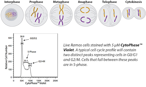
  • Additional reagents for proliferation, long-term and short-term cell tracking, cell cycle analysis, and mitochondrial staining.

Live/Dead Indicators

Within a sample, it can be important to assess the viability and general health of a cell. Dying/dead cells can create debris that lead to false positive staining within a flow cytometry experiment. A majority of the reagents in this category function on the concept that intact membranes of living cells help to limit the binding of a fluorescent dye or chemical probe. Learn about each category of our non-antibody chemical probes below to help decide which reagent is ideal for your assay.

Fixable Live/Dead Indicators

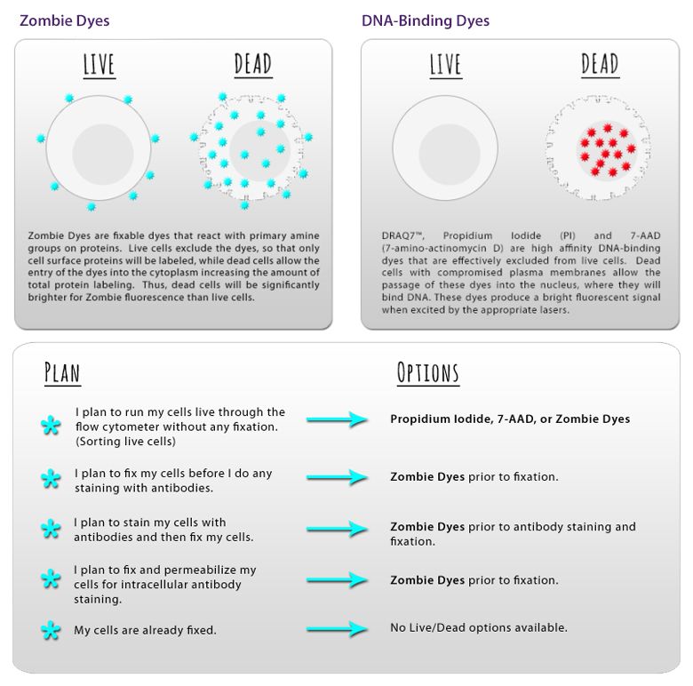
Fixation and permeabilization reagents (such as alcohols and paraformaldehyde) can unwind and denature DNA, dislodging DNA-binding dyes and giving false negative results. Simultaneously, free dye could find its way into previously healthy cells that have now been fixed/permeabilized and give a false positive result.


To avoid this issue, we provide Zombie Dyes, which bind the amine groups present on proteins. While live cells are stained to a low degree due to surface proteins, dead cells will stain much more brightly due to the high abundance of internal proteins that can be stained once membranes are compromised.


Learn about Zombie Dyes >

Non-Fixable Live/Dead Indicators

One of the earliest techniques for identifying dead cells involved the use of nucleic acid-binding dyes such as Propidium Iodide, 7-AAD, DAPI, and BioLegend’s Helix NP™ (non-permeant) dyes. These dyes work on the principle that live and healthy cells possess intact membranes that prevent these dyes from gaining access to the nucleus. However, dying cells will have compromised membranes that allow these dyes to bind directly to the DNA.


It should be noted that fixation and permeabilization procedures can cause potential issues with the ability of these dyes to remain bound to their target.


View Our DNA-Binding Dyes >

Live/Dead Indicators

Within a sample, it can be important to assess the viability and general health of a cell. Dying/dead cells can create debris that lead to false positive staining within a flow cytometry experiment. A majority of the reagents in this category function on the concept that intact membranes of living cells help to limit the binding of a fluorescent dye or chemical probe. Learn about each category of our non-antibody chemical probes below to help decide which reagent is ideal for your assay.

Fixable Live/Dead Indicators

Fixation and permeabilization reagents (such as alcohols and paraformaldehyde) can unwind and denature DNA, dislodging DNA-binding dyes and giving false negative results. Simultaneously, free dye could find its way into previously healthy cells that have now been fixed/permeabilized and give a false positive result.


To avoid this issue, we provide Zombie Dyes, which bind the amine groups present on proteins. While live cells are stained to a low degree due to surface proteins, dead cells will stain much more brightly due to the high abundance of internal proteins that can be stained once membranes are compromised.


Learn about Zombie Dyes >

Non-Fixable Live/Dead Indicators

One of the earliest techniques for identifying dead cells involved the use of nucleic acid-binding dyes such as Propidium Iodide, 7-AAD, DAPI, and BioLegend’s Helix NP™ (non-permeant) dyes. These dyes work on the principle that live and healthy cells possess intact membranes that prevent these dyes from gaining access to the nucleus. However, dying cells will have compromised membranes that allow these dyes to bind directly to the DNA.


It should be noted that fixation and permeabilization procedures can cause potential issues with the ability of these dyes to remain bound to their target.


View our DNA-binding dyes >

Apoptosis Indicators

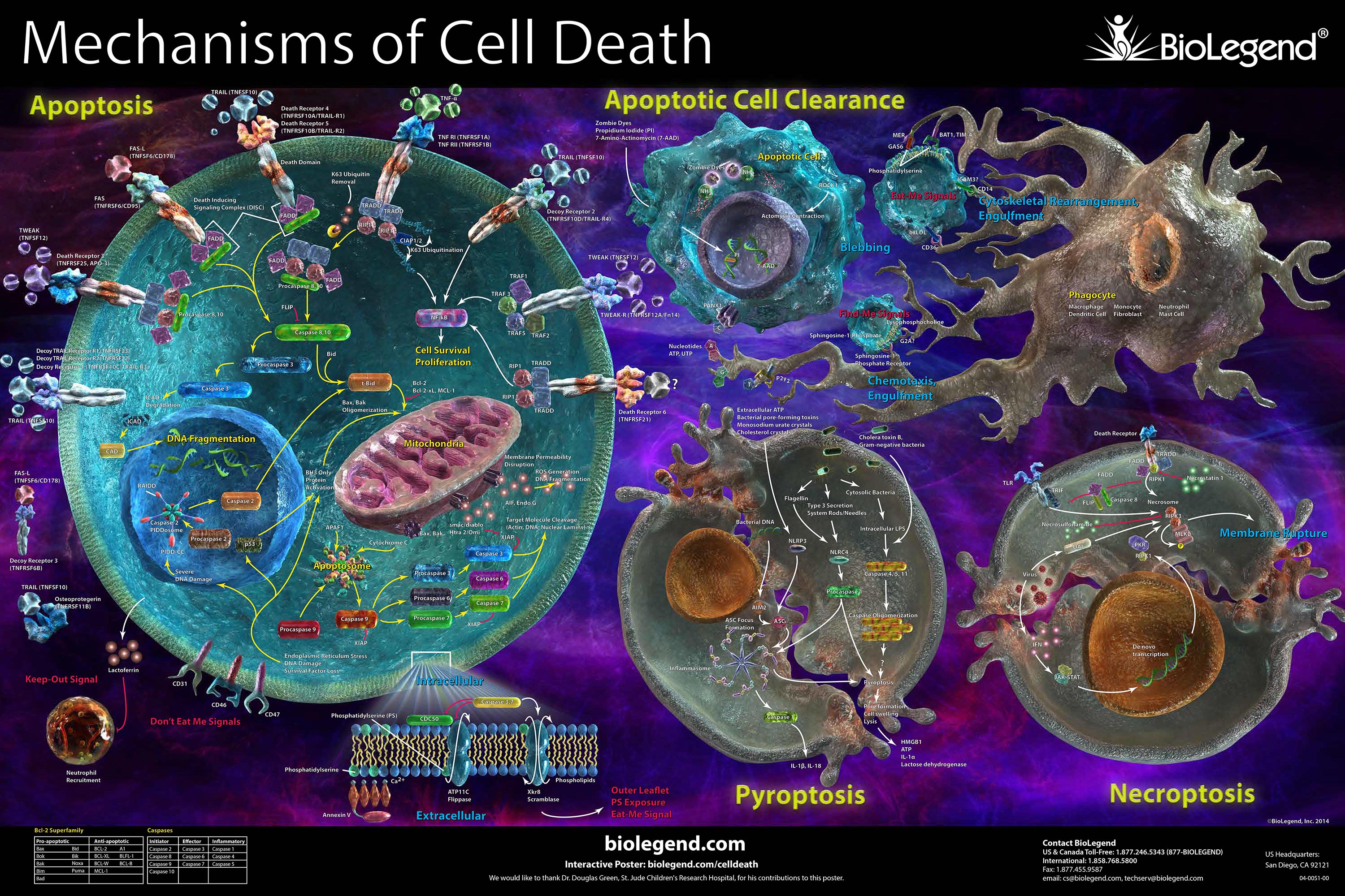
Apoptosis is a type of programmed cell death used by cells for self-elimination. Apoptotic cells do not release their cellular contents and are quickly engulfed by phagocytic cells, and therefore avoid triggering unnecessary inflammatory responses. Since many physiological functions rely on the proper elimination of cells, dysregulation of apoptosis can contribute to diseases like cancer and neurodegeneration. You can learn about some of the chemical probes we offer for the initial stages of apoptosis identification or utilize our apoptosis webpage for Caspase, Death Receptor, and Ligand reagents.


View our apoptosis webpage >


Calcium-Independent Probes

The addition of calcium to some samples may affect the viability and marker expression of cell types. For scientists looking to use calcium-independent probes that do not require a specialized buffer, we provide Apotracker™. Apotracker™ is a family of fluorogenic probes that bind to apoptotic cells in a calcium-independent manner, exhibiting a linear relationship with Annexin V staining which suggests they are both detecting externalized phosphatidylserine (PS) residues. In addition, Apotracker™ can be useful for microscopy assays where it presents less background staining than Annexin V.


Learn more about Apotracker™ >


Calcium-Dependent Probes

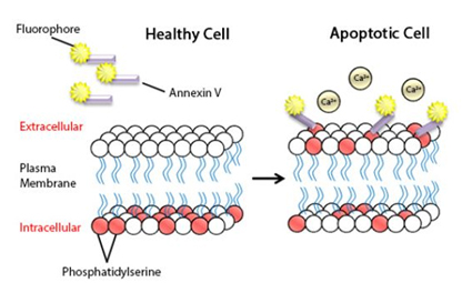
Annexin V is one of the most common probes used to examine early-to-mid stages of apoptosis. Annexin V is a protein that recognizes phosphatidylserine (PS), which is commonly found on the cytosolic side of the membrane in healthy cells. However, in dying or apoptotic cells, PS translocates to the extracellular side of the membrane, exposing it and making it available for binding by Annexin V in a calcium-dependent manner. BioLegend provides several fluorophore-conjugated Annexin V options, an array of impermeant nucleic acid stains to be used for distinguishing apoptotic from necrotic cells, and the Annexin V Binding Buffer required for the calcium-dependent binding of the annexin to PS.


View our Annexin V reagents >

Additional Chemical Probes

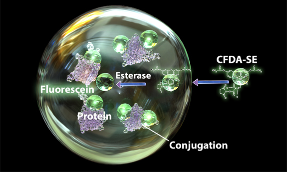
In addition to our chemical probes dedicated to identifying live vs. dead cells, we offer additional dyes that can be utilized to understand various aspects of a cell’s health. This includes the assessment of the proliferation status of cells through short and long-term trackers, cell cycle analysis tools, and vitality indicators such as esterase-dependent probes and mitochondria-reactive dyes.


View all cell health chemical probes >


Cell Cycle Reagents

Cells can divide in response to stimuli such as growth factors and cytokines, or specific antigens/mitogens. This response must be tightly regulated as improper cell proliferation can lead to tumor growth or developmental problems. Browse through our antibodies for importnat cell cycle progression proteins like cyclins, bioactive and stable recombinant proteins for stimulating or suppressing growth, and specialized dyes for tracking cell proliferation.


View our cell cycle reagents >


Dead Cell Removal

Dead cells or debris can often interfere with obtaining reliable readouts. Dead cells in particular can bind to antibodies non-specifically and, in the case of multiomics applications, take up precious reads in a library. The reagents listed below provide options for researchers to remove dead cells from their samples for downstream analysis such as single-cell sequencing, cell line development, and flow cytometry.


MojoSort™ Human and Mouse Dead Cell Removal Kits


Dead cells are magnetically separated in standard cell separation buffer containing no calcium (MojoSort™ Buffer, or equivalent), resulting in high live cell enrichment and superior yield and cell recovery.


View our MojoSort™ Dead Cell Removal Kits >


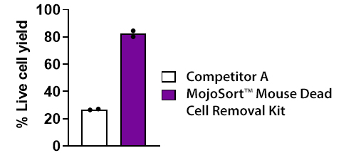
Representative live cell recovery comparison between competitor product A and MojoSort™ Mouse Dead Cell Removal Kit. Dead cells were magnetically separated from heat-stressed C57BL/6 murine splenocytes (containing 40% dead cells) using recommended protocols for each kit. Competitor A required use of a specialized binding buffer containing high Ca2+ concentration. % live cell yield was calculated based on recovery of CD45+ Helix NP™ Blue- Apotracker™ Green- population relative to the unsorted population. Data represents compiled data from two technical replicates.


Application data: Live cell enrichment using MojoSort™ Human Dead Cell Removal Kit for scRNA-seq and CITE-seq


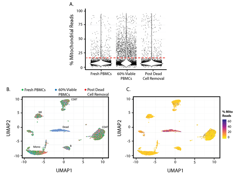
Live cell enriched samples can be used in a variety of downstream applications, where cell viability is critical for generating high-quality results. This includes single-cell RNA sequencing (scRNA-seq) and associated epitope detection using Cellular Indexing of Transcriptomics and Epitomics (CITE-seq) using our TotalSeq™ oligonucleotide conjugated antibodies. 


Three cell samples were collected and pooled on one CITE-seq experiment: Fresh, Human PBMCs from a healthy donor; 60% Viable PBMCs, a cell pool that contains 60% live PBMCs; and Post Dead Cell Removal, which contains the 60% Viable PBMCs pool cleaned up by MojoSort™ Human Dead Cell Removal Kit. A) Cells with higher Percent Mitochondrial Read in a library are commonly considered to have low cell viability. These dead and dying cells are abundant in the 60% Viable PBMCs, group, and can be removed by MojoSort™ Human Dead Cell Removal Kit. B) UMAP based on TBNK lineage protein marker expression showed common PBMC cell type can be detected from all three samples, with the exception of a dead cell cluster in the center that consists of mostly cells from the partial dead cell group 60% Viable PBMCs. C) The same UMAP that colored by Percent Mitochondrial Reads per cell indicates the center cell cluster are dead and dying cells.


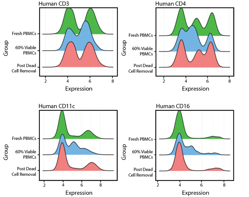
Ridge plots showed that the MojoSort™ Human Dead Cell Removal Kit preserve all common lineages in PBMCs, and the expression levels of the lineage markers were mostly the same when comparing the Post Dead Cell Removal group to the Fresh PBMCs group.

ProductsHere

Login / Register
Remember me
Forgot your password? Reset password?
Create an Account