- Regulatory Status
- RUO
- Other Names
- Clearing-enhanced 3D
- Ave. Rating
- Submit a Review
- Product Citations
- publications

-

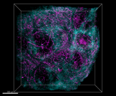
Paraformaldehyde-fixed (4%), 500 µm-thick mouse spleen section was processed according to the Ce3D™ Tissue Clearing Kit protocol (Cat. No. 480401). The section was anti-mouse CD172a (SIRPα) (clone P84) Alexa Fluor® 594 at 5 µg/mL (cyan), and anti-mouse CD200 (OX2) (clone OX-90) Alexa Fluor® 647 at 5 µlg/mL (magenta). The section was then optically cleared and mounted in a sample chamber. The image was captured with a 10X objective using Zeiss 780 confocal microscope and processed by Imaris image analysis software. Watch the video. -

Paraformaldehyde-fixed (4%), 500 µm-thick mouse spleen section was processed according to the Ce3D™ Tissue Clearing Kit protocol (Cat. No. 480401). The section was costained with anti-mouse CD169 (Siglec-1) (clone 3D6.112) Alexa Fluor® 488 at 5 µg/mL (green), anti-mouse I-A/I-E (clone M5/114.15.2) Alexa Fluor® 594 at 5 µg/mL (blue), and anti-mouse CD3ε (clone KT3.1.1) Alexa Fluor® 647 at 5 µg/mL (magenta). The section was then optically cleared and mounted in a sample chamber. The image was captured with a 10X objective using Zeiss 780 confocal microscope and processed by Imaris image analysis software. Watch the video. -

Paraformaldehyde-fixed (4%), mouse intestine section was processed according to the Ce3D™ Tissue Clearing Kit protocol (Cat. No. 480401). The section was costained with anti-Tubulin β 3 (TUBB3) (clone TUJ1) Alexa Fluor® 594 at 5 µg/mL (green), and anti-mouse CD45 (clone 30-F11) Alexa Fluor® 647 at 5 µg/mL (magenta). The section was then optically cleared and mounted in a sample chamber. The image was captured with a 20X objective using Zeiss 780 confocal microscope and processed by Imaris image analysis software. Watch the video.
| Cat # | Size | Price | Quantity Check Availability | Save | ||
|---|---|---|---|---|---|---|
| 427701 | 10 tests | 244€ | ||||
| 427702 | 50 tests | 620€ | ||||
Ce3D™ (clearing-enhanced 3D) Tissue Clearing Solution is a hydrophilic, refractive index (RI) matching reagent that is easy-to-use, and allows rapid and robust clearing of diverse tissues. Ce3D™ has an RI of ~1.5 and is compatible with multicolor fluorescence microscopy. The Ce3D™ tissue clearing technique works by simple immersion of tissue into the clearing reagent, and does not require special equipment or containers. Ce3D™ Tissue Clearing Kit includes Ce3D™ Tissue Clearing Solution, Ce3D™ Permeabilization/Blocking Buffer, Ce3D™ Antibody Diluent Buffer and Ce3D™ Wash Buffer, which are formulated for use with 3D IHC application.
Product DetailsProduct Details
- Storage & Handling
- The buffer solution should be stored between 2°C and 8°C.
- Application
-
IHC-F - Quality tested
3D IHC - Verified - Recommended Usage
-
Ce3D™ tissue clearing solution is used at 500 µL per test size (500 µm thick tissue) to cover the tissue.
Ce3D™ Permeabilization/blocking buffer is used at 500 µL per test size (500 µm thick tissue) to cover the tissue.
Ce3D™ antibody diluent buffer is used at 500 µL per test size (500 µm thick tissue) to cover the tissue.
Ce3D™ wash buffer is used at 500 µL per test size (500 µm thick tissue) to cover the tissue. - Product Citations
-
Antigen Details
- Gene ID
- NA
- App Abbreviation (DOES NOT SHOW ON TDS):
- IHC-F,3D IHC

 Login / Register


Follow Us