- Clone
- M380B (See other available formats)
- Regulatory Status
- RUO
- Isotype
- Mouse IgG1
- Ave. Rating
- Submit a Review
- Product Citations
- publications

-

PBMC untreated (negative control, open histogram) or treated with cell activation cocktail (Cat No. 423301) (positive control, solid histogram) were fixed and permeabilized using True Phos™ Perm Buffer (Cat No. 425401) and then intracellularly stained with either Alexa Fluor® 647 anti-Phosphoserine (clone M380B) or Alexa Fluor® 647 Mouse IgG1, κ Isotype Control (Cat No. 400155) (open histogram, dashed line)(Representative for untreated and treated cells).
| Cat # | Size | Price | Quantity Check Availability | Save | ||
|---|---|---|---|---|---|---|
| 944103 | 25 tests | 144€ | ||||
| 944104 | 100 tests | 320€ | ||||
Phosphorylation of specific serine residues is a post-translational modification event critical for regulating the activity of many proteins. Protein phosphorylation events can alter protein function through a variety of molecular mechanisms, ranging from conformational changes that can stimulate or inhibit protein function to facilitating protein-protein interactions with binding partners. Antibodies that can detect phosphoserine residues are valuable tools for identifying novel phospho-sites and characterizing changes in the post-translational state of a broad range of phosphorylated proteins.
Product DetailsProduct Details
- Verified Reactivity
- Human, Mouse, Rat, All Species
- Antibody Type
- Monoclonal
- Host Species
- Mouse
- Immunogen
- Synthetic peptide containing phosphorylated serine
- Formulation
- Phosphate-buffered solution, pH 7.2, containing 0.09% sodium azide and BSA (origin USA)
- Preparation
- The antibody was purified by affinity chromatography and conjugated with Alexa Fluor® 647 under optimal conditions.
- Concentration
- Lot-specific (to obtain lot-specific concentration and expiration, please enter the lot number in our Certificate of Analysis online tool.)
- Storage & Handling
- The antibody solution should be stored undiluted between 2°C and 8°C, and protected from prolonged exposure to light. Do not freeze.
- Application
-
ICFC - Quality tested
- Recommended Usage
-
Each lot of this antibody is quality control tested by intracellular immunofluorescent staining with flow cytometric analysis. For flow cytometric staining, the suggested use of this reagent is 5 µL per million cells in 100 µL staining volume or 5 µL per 100 µL of whole blood. It is recommended that the reagent be titrated for optimal performance for each application.
* Alexa Fluor® 647 has a maximum emission of 668 nm when it is excited at 633 nm / 635 nm.
Alexa Fluor® and Pacific Blue™ are trademarks of Life Technologies Corporation.
View full statement regarding label licenses - Excitation Laser
-
Red Laser (633 nm)
Antigen Details
- Function
- Cell signaling
- Biology Area
- Cell Biology, Immunology, Signal Transduction
- Molecular Family
- Phospho-Proteins, Protein Kinases/Phosphatase
- Antigen References
-
- Rogerson D. et. al. 2015. Nat. Chem. Biol. 11:496-503.
- Steinfield J. et. al. 2014. ACS Chem. Biol. 9(5):1104-1112.
- Gene ID
- NA
- UniProt
- View information about Phosphoserine on UniProt.org
Related FAQs
Other Formats
View All Phosphoserine Reagents Request Custom Conjugation| Description | Clone | Applications |
|---|---|---|
| Purified anti-Phosphoserine | M380B | WB,ICC,IP,ICFC |
| Alexa Fluor® 647 anti-Phosphoserine Antibody | M380B | ICFC |
Compare Data Across All Formats
This data display is provided for general comparisons between formats.
Your actual data may vary due to variations in samples, target cells, instruments and their settings, staining conditions, and other factors.
If you need assistance with selecting the best format contact our expert technical support team.
-
Purified anti-Phosphoserine

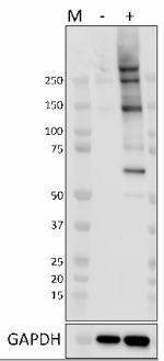
Whole cell lysates (15 μg) from untreated (-) or Calyculin A... 
HeLa cells were fixed with 4% paraformaldehyde for 10 minute... 
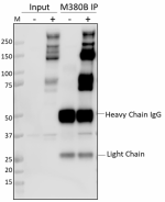
Whole cell extracts (250 µg total protein) prepared from HeL... 
PBMCs treated with (positive control, filled histogram) or w... -
Alexa Fluor® 647 anti-Phosphoserine Antibody

PBMC untreated (negative control, open histogram) or treated...
 Login / Register


Follow Us