- Regulatory Status
- RUO
- Other Names
- T-Cell Leukemia Antigen, gp40, TP41, T-Cell Surface Antigen Leu-9, LEU-9, P41 Protein, Tp40, T-Cell Antigen CD7
- Ave. Rating
- Submit a Review
- Product Citations
- publications

-

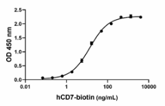
Biotinylated recombinant human CD7 binds to immobilized recombinant human SECTM1 in a dose-dependent manner.The ED50 for this effect is 7 – 35 ng/mL. -

Stability Testing for Biotinylated Recombinant Human CD7. Biotinylated recombinant human CD7 was aliquoted in PBS, pH 7.2. One aliquot was frozen and thawed four times (4x Freeze/Thaw) and compared to the control that was kept at 4°C (Control). The samples were tested for their ability to bind to immobilized recombinant human SECTM1 in a dose-dependent manner. The ED50 for this effect is 7 – 35 ng/mL.
| Cat # | Size | Price | Quantity Check Availability | Save | ||
|---|---|---|---|---|---|---|
| 796304 | 25 µg | 179€ | ||||
| 796306 | 100 µg | 423€ | ||||
The CD7 is type I transmembrane glycoprotein with a MW of 40-kD (gp40) and contains a single-domain Ig; thus, CD7 is a Ig superfamily member. It is expressed on T cell precursors, T cells, natural killer (NK) cells, myeloid precursor cells, plasmacytoid dendritic cells (DCs), pre-B cells, and in leukemia cells. Human CD7 is a protein of 240 amino acids and the mature protein includes amino acid 26 to 240. The extracellular domain contains residues 26 to 180. The intracellular domain possesses a YEEM motif, like CD28. YEEM motif activates the PI3K signaling pathway and the treatment of cells with anti-CD3 or PMA in addition to anti-CD7 showed that CD7 is mitogenic for human T cells and induces IL-2. It has been described a C7-, CD4+ subpopulation of memory T cells that specially accumulate in skin lesions associated to chronic inflammatory conditions. CD7 is overexpressed in classical Hodgkin lymphoma‐infiltrating T lymphocyte. CD7 is also expressed in T-cell acute lymphoblastic leukemia (T-ALL). CD7 has been used as a novel approach based on chimeric antigen receptor (CAR)–redirected T lymphocytes.
Product DetailsProduct Details
- Source
- Human CD7, amino acids (Ala26-Pro180) (Accession No. P09564) with a linker, a C-terminal GGS-8His and an Avi-tag, was expressed in 293E cells. Human CD7-Avi tag was site-specifically biotinylated by enzyme BirA.
- Molecular Mass
- The 192 amino acid recombinant protein has a predicted molecular mass of approximately 20.2 KD. The DTT-reduced and non-reduced glycosylated protein migrate at approximately 30 - 40 kD and 27 - 40 kD respectively by SDS-PAGE. The predicted N-terminal amino acid is Ala.
- Purity
- > 95%, as determined by Coomassie stained SDS-PAGE
- Formulation
- 0.22 µm filtered protein solution is PBS, pH 7.2
- Endotoxin Level
- Less than 0.1 EU per µg cytokine as determined by the LAL method
- Concentration
- 25 µg size is bottled at 200 µg/mL. 100 µg size and larger sizes are lot-specific and bottled at the concentration indicated on the vial. To obtain lot-specific concentration and expiration, please enter the lot number in our Certificate of Analysis online tool.
- Storage & Handling
- Unopened vial can be stored between 2°C and 8°C for up to 2 weeks at -20°C for up to six months, or at -70°C or colder until the expiration date. For maximum results, quick spin vial prior to opening. The protein can be aliquoted and stored at -20°C or colder. Stock solutions can also be prepared at 50 - 100 µg/mL in appropriate sterile buffer, carrier protein such as 0.2 - 1% BSA or HSA can be added when preparing the stock solution. Aliquots can be stored between 2°C and 8°C for up to one week and stored at -20°C or colder for up to 3 months. Avoid repeated freeze/thaw cycles.
- Activity
- Biotinylated recombinant human CD7 binds to immobilized recombinant human SECTM1 in a dose-dependent manner. The ED50 for this effect is 7 – 35 ng/mL.
- Application
-
Bioassay
- Application Notes
-
BioLegend carrier-free recombinant proteins provided in liquid format are shipped on blue ice. Our comparison testing data indicates that when handled and stored as recommended, the liquid format has equal or better stability and shelf-life compared to commercially available lyophilized proteins after reconstitution. Our liquid proteins are validated in-house to maintain activity after shipping on blue ice and are backed by our 100% satisfaction guarantee. If you have any concerns, contact us at tech@biolegend.com.
Antigen Details
- Distribution
-
T cells, NK cells, early stages of T cells, myeloid cell precursors, and CD19+ B progenitor cells. Highly expressed on malignant immature T cells. Expressed in monocytes and macrophages
- Function
- Regulation of peripheral T and NK cell cytokine production, functions as a costimulatory receptor for T cell proliferation. CD7 is upregulated in CD4 and CD8 T cells stimulated with anti-CD3 plus anti-CD28.
- Interaction
- CD7 interacts with its ligand SECTM1 in soluble form which is highly expressed by cancer cells.
- Ligand/Receptor
- SECTM1, CTSL
- Bioactivity
- Biotinylated recombinant human CD7 binds to immobilized recombinant human SECTM1 in a dose-dependent manner.
- Cell Type
- Dendritic cells, Macrophages, Monocytes, NK cells, T cells, Thymocytes, Tregs
- Biology Area
- Adaptive Immunity, Cancer Biomarkers, Cell Biology, Cell Proliferation and Viability, Costimulatory Molecules, Immuno-Oncology
- Molecular Family
- CD Molecules
- Antigen References
-
- Lyman SD, et al., 2000. J. Bio. Chem. 275: P3431-7.
- L Liu, et al., 2000. Clin. Exp. Immunol 121:94-9.
- Milush JM, et al., 2009. Blood 114:4823-31.
- Seegmiller AC, et al., 2009. Cytometry B Clin Cytom. 76:169-74.
- Wang T, et al., 2012. J Leukoc Biol 91: 449-59.
- Wang T, et al., 2014. J Invest Dermatol 134:1108-1118.
- Png YT, et al., 2017. Blood Adv 1: 2348-60.
- Haftcheshmeh SM 2019. J Cell Physiol 234:1179-89.
- Gene ID
- 924 View all products for this Gene ID
- UniProt
- View information about CD7 on UniProt.org

 Login / Register


Follow Us