Analyte Specific Reagent. Analytical and performance characteristics are not established.
- Clone
- NAT105
- Other Names
- PD1
- Isotype
- Mouse IgG1, κ
-

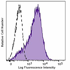
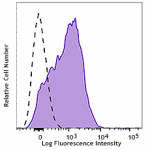
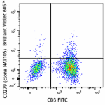
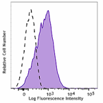
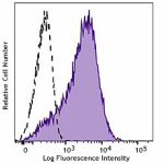
Typical results from PHA-stimulated human peripheral blood lymphocytes stained either with NAT105 PE used at 5 μL/test (filled histogram) or with an isotype control (open histogram).
| Cat # | Size | Price | Quantity Check Availability | Save | ||
|---|---|---|---|---|---|---|
| 981602 | 500 µL | 258€ | ||||
CD279, also known as Programmed cell death 1 (PD-1), is a 55 kD member of the immunoglobulin superfamily. CD279 contains the immunoreceptor tyrosine-based inhibitory motif (ITIM) in the cytoplasmic region and plays a key role in peripheral tolerance and autoimmune diseases. CD279 is expressed predominantly on activated T cells, B cells, and myeloid cells. PD-L1 (B7-H1, CD274) and PD-L2 (B7-DC, CD273) are ligands of CD279 (PD-1) and are members of the B7 gene family. Evidence suggests overlapping functions for these two PD-1 ligands and their constitutive expression on some normal tissues and upregulation on activated antigen-presenting cells. The interaction with CD279 ligands results in inhibition of T cell proliferation and cytokine secretion.
Product DetailsProduct Details
- Reactivity
- Human
- Formulation
- Phosphate-buffered solution, pH7.2, containing 0.09% sodium azide, 0.2% (w/v) BSA (origin USA), and a stabilizer.
- Preparation
- The antibody was purified by affinity chromatography and conjugated with PE under optimal conditions.
- Concentration
- 100 µg/mL
- Storage & Handling
- The antibody solution should be stored undiluted between 2°C and 8°C, and protected from prolonged exposure to light. Do not freeze.
- Application
-
Suggested for Flow Cytometry
- Disclaimer
-
WARNINGS AND PRECAUTIONS
- Use appropriate personal protective equipment and safety practices per universal precautions when working with this reagent. Refer to the reagent safety data sheet.
- This antibody contains sodium azide. Follow federal, state and local regulations to dispose of this reagent. Sodium azide build-up in metal wastepipes may lead to explosive conditions; if disposing of reagent down wastepipes, flush with water after disposal.
- All specimens, samples and any material coming in contact with them should be considered potentially infectious and should be disposed of with proper precautions and in accordance with federal, state and local regulations.
- Do not use this reagent beyond the expiration date stated on the label.
- Do not use this reagent if it appears cloudy or if there is any change in the appearance of the reagent as these may be an indication of possible deterioration.
- Avoid prolonged exposure of the reagent or stained cells to light.
Antigen Details
- Antigen References
-
- Francisco LM, et al. 2010. Immunological Rev. 236:219.
Compare Data Across All Formats
This data display is provided for general comparisons between formats.
Your actual data may vary due to variations in samples, target cells, instruments and their settings, staining conditions, and other factors.
If you need assistance with selecting the best format contact our expert technical support team.
-
Purified anti-human CD279 (PD-1)
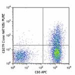
Human peripheral blood lymphocytes were stained with purifie... 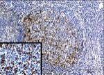
Tissue imaged here is the lymphoid nodule/germinal center of... 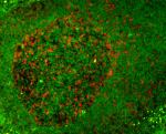
Human paraffin-embedded tonsil tissue slices were prepared w... 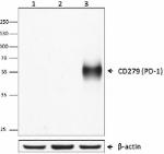
Western blot analysis of Jurkat (lane 1), PBMC untreated (la... 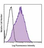
PHA-stimulated (three days) human PBMC were stained with pur... -
PE anti-human CD279 (PD-1)
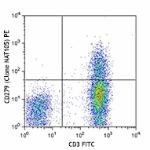
Human peripheral blood lymphocytes were stained with CD3 FIT... 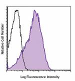
PHA-stimulated (three days) human PBMC were stained with CD2... 
Multiplexed IHC staining of PE anti-CD279 (clone NAT105) on ... -
APC anti-human CD279 (PD-1)
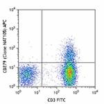
Human peripheral blood lymphocytes were stained with CD3 FIT... 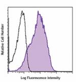
PHA-stimulated (three days) human PBMC were stained with CD2... -
Alexa Fluor® 488 anti-human CD279 (PD-1)
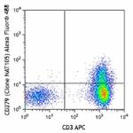
Human peripheral blood lymphocytes were stained with CD3 APC... 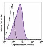
PHA-stimulated (3-day) human peripheral blood lymphocytes we... -
PerCP/Cyanine5.5 anti-human CD279 (PD-1)
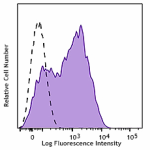
PHA-stimulated (3-day) human peripheral blood mononuclear ce... 
Human peripheral blood lymphocytes were stained with CD3 APC... -
PE/Cyanine7 anti-human CD279 (PD-1)
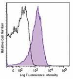
PHA-stimulated (3-day) human peripheral blood lymphocytes we... -
FITC anti-human CD279 (PD-1)
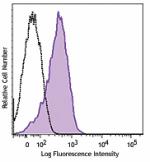
PHA-stimulated (3-day) human peripheral blood lymphocytes we... -
APC/Cyanine7 anti-human CD279 (PD-1)
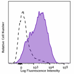
PHA-stimulated (3-day) human peripheral blood lymphocytes we... -
Alexa Fluor® 647 anti-human CD279 (PD-1)
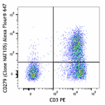
Human peripheral blood lymphocytes were stained with CD3 PE ... 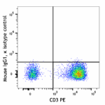
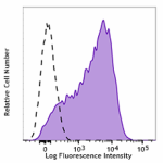
PHA-stimulated (3-day) human peripheral blood lymphocytes we... -
Biotin anti-human CD279 (PD-1)
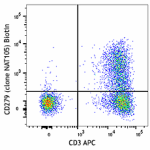
Human peripheral blood lymphocytes were stained with CD3 APC... 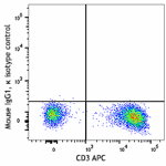
-
Brilliant Violet 510™ anti-human CD279 (PD-1)
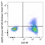
Human peripheral blood lymphocytes were stained with CD3 PE ... 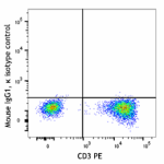
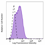
PHA-stimulated (3-day) human peripheral blood lymphocytes we... -
Brilliant Violet 421™ anti-human CD279 (PD-1)
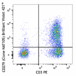
Human peripheral blood lymphocytes were stained with CD3 PE ... 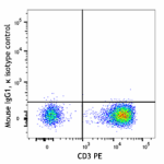
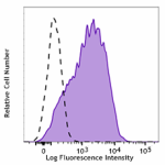
PHA-stimulated (3-day) human peripheral blood lymphocytes we... -
Brilliant Violet 711™ anti-human CD279 (PD-1)
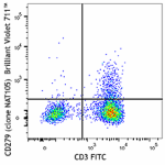
Human peripheral blood lymphocytes were stained with CD3 FIT... 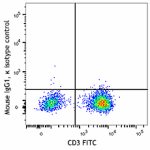
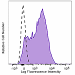
PHA-stimulated (3-day) human peripheral blood lymphocytes we... -
Brilliant Violet 785™ anti-human CD279 (PD-1)
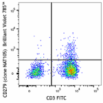
Human peripheral blood lymphocytes were stained with CD3 FIT... 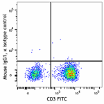
PHA-stimulated (3-day) human peripheral blood lymphocytes we... -
Brilliant Violet 605™ anti-human CD279 (PD-1)
Human peripheral blood lymphocytes were stained with CD3 FIT... 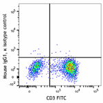
PHA-stimulated (3-day) human peripheral blood lymphocytes we... -
Brilliant Violet 650™ anti-human CD279 (PD-1)

Human peripheral blood lymphocytes were stained with CD3 FIT... 
PHA-stimulated (3-day) human peripheral blood lymphocytes we... -
PE/Dazzle™ 594 anti-human CD279 (PD-1)

Human peripheral blood lymphocytes were stained with CD3 APC... -
TotalSeq™-Bn1303 anti-human CD279 (PD-1)
-
PE anti-human CD279
Typical results from PHA-stimulated human peripheral blood l... -
Spark Red™ 718 anti-human CD279 (PD-1) (Flexi-Fluor™)
-
APC anti-human CD279

Typical results from PHA-stimulated human peripheral blood l...
 Login / Register


Follow Us