- Clone
- mN1A (See other available formats)
- Regulatory Status
- RUO
- Other Names
- Neurogenic locus notch homolog protein 1, Notch-1
- Isotype
- Mouse IgG1, κ
- Ave. Rating
- Submit a Review
- Product Citations
- 2 publications
| Cat # | Size | Price | Quantity Check Availability | Save | ||
|---|---|---|---|---|---|---|
| 629105 | 25 µg | 84€ | ||||
| 629106 | 100 µg | 162€ | ||||
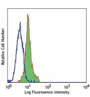
Notch1, also known as neurogenic locus notch homolog protein 1, is a large >270 kD protein that functions as a receptor for the membrane ligands Jagged1, Jagged2 and Delta1 to regulate cell fate decisions. Upon ligand activation, the transmembrane Notch1 receptor is cleaved by TNF-alpha converting enzyme (TACE) to produce a membrane-associated intermediate fragment (NEXT). This fragment is further cleaved by presenilin-dependent gamma secretase to release a notch-derived peptide containing the intracellular fragment from the membrane. The released Notch intracellular fragment (NICD) translocates to the nucleus and forms a transcriptional complex with the RBP-J κ transcriptional activator complex to alter differentiation, proliferation, and apoptotic programs. Notch 1 is highly expressed in the brain, lung, and thymus (CD4-CD8- cells and CD4-CD8+ cells) with lower levels of expression observed in the spleen, bone marrow, spinal cord, eyes, mammary gland, liver, intestine, kidney and heart. The transmembrane Notch protein is a heterodimeric protein consisting of a C-terminal fragment and N-terminal fragment (probably linked by disulphide bonds) containing 5 ankyrin repeats, 36 EGF repeats, and 3 Notch/Lin repeats. Notch1 can be modified by phosphorylation. The mN1A monoclonal antibody reacts with the intracellular domain of mouse and human Notch1 and has been reported to have highest affinity for activated intracellular Notch1 and lower affinity for full-length unprocessed/heterodimeric Notch1 forms. This antibody does not recognize rat Notch1 or cross-react with Notch2, 3, or 4.
Product DetailsProduct Details
- Verified Reactivity
- Mouse, Human
- Antibody Type
- Monoclonal
- Host Species
- Mouse
- Immunogen
- Notch1 GST fusion protein
- Formulation
- Phosphate-buffered solution, pH 7.2, containing 0.09% sodium azide.
- Preparation
- The antibody was purified by affinity chromatography, and conjugated with PE under optimal conditions.
- Concentration
- 0.2 mg/ml
- Storage & Handling
- The antibody solution should be stored undiluted between 2°C and 8°C, and protected from prolonged exposure to light. Do not freeze.
- Application
-
ICFC - Quality tested
- Recommended Usage
-
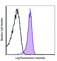
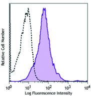
Each lot of this antibody is quality control tested by intracellular immunofluorescent staining with flow cytometric analysis. For flow cytometric staining, the suggested use of this reagent is ≤1.0 µg per million cells in 100 µl volume. It is recommended that the reagent be titrated for optimal performance for each application.
- Excitation Laser
-
Blue Laser (488 nm)
Green Laser (532 nm)/Yellow-Green Laser (561 nm)
- Application Notes
-
Additional reported applications of this antibody (for the relevant formats) include: Western blotting1,2,3, and immunohistochemistry4,5.
-
Application References
(PubMed link indicates BioLegend citation) -
- Huppert SS, et al. 2000. Nature 405:966. (WB)
- Ray WJ, et al. 1999. Proc. Natl. Acad. Sci. USA 96:3263. (WB)
- DeStrooper B, et al. 1999. Nature 398:518. (WB)
- Sun H, et al. 2007. J. Cell Biol. 177:647. (IHC) PubMed
- Jundt F, et al. 2008. Leukemia. 22:1587. (IHC) PubMed
- Liu JC, et al. 2012. PNAS 109:5832. PubMed.
- Product Citations
-
- RRID
-
AB_2282905 (BioLegend Cat. No. 629105)
AB_2282905 (BioLegend Cat. No. 629106)
Antigen Details
- Structure
- Transmembrane receptor, heterodimer consisting of a C-terminal fragment and N-terminal fragment probably linked by disulphide bonds. Contains 5 ankyrin repeats, 36 EGF repeats, 3 Notch/Lin repeats. Predicted molecular weight 271 kD.
- Distribution
-
Highly expressed in the brain, lung, and thymus (CD4-CD8- cells and CD4-CD8+ cells). Lower levels of expression in spleen, bone marrow, spinal cord, eyes, mammary gland, liver, intestine, kidney and heart.
- Function
- Receptor for the membrane ligands Jagged1, Jagged2 and Delta1 to regulate cell fate decisions.
- Modification
- Phosphorylation
- Cell Type
- Neural Stem Cells, T cells
- Biology Area
- Cell Biology, Immunology, Neuroscience, Neuroscience Cell Markers, Stem Cells, Synaptic Biology
- Molecular Family
- Postsynaptic proteins
- Antigen References
-
1. Huppert SS, et al. 2000. Nature 405:966.
2. Kopan R, et al. 1993. J. Cell Biol. 121:631.
3. Saxena MT, et al. 2001. J. Biol. Chem. 276:40268.
4. Mizutani T, et al. 2001. Proc. Natl. Acad. Sci. USA 98:9026. - Gene ID
- 4851 View all products for this Gene ID
- UniProt
- View information about Notch 1 on UniProt.org
Related FAQs
- What type of PE do you use in your conjugates?
- We use R-PE in our conjugates.
Other Formats
View All Notch 1 Reagents Request Custom Conjugation| Description | Clone | Applications |
|---|---|---|
| PE anti-Notch 1 | mN1A | ICFC |
Customers Also Purchased
Compare Data Across All Formats
This data display is provided for general comparisons between formats.
Your actual data may vary due to variations in samples, target cells, instruments and their settings, staining conditions, and other factors.
If you need assistance with selecting the best format contact our expert technical support team.
-
PE anti-Notch 1
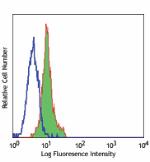
Jurkat cells Intracellularly stained with Notch-1 PE


 Login / Register




Follow Us