 Login / Register


- Clone
- A20017A (See other available formats)
- Regulatory Status
- RUO
- Other Names
- PRKAA1, PRKAA2, AMPKa1, AMPKa2, AMPK, ACACA Kinase, Tau-Protein Kinase PRKAA1, Tau-Protein Kinase PRKAA2, Protein kinase AMP-Activated, Alpha 1 Catalytic Subunit, HMGCR Kinase
- Isotype
- Mouse IgG1, κ
- Ave. Rating
- Submit a Review
- Product Citations
- publications

-

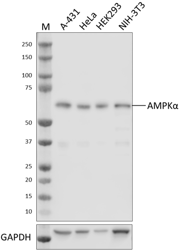
Whole cell extracts (15 µg total protein) from serum-starved HEK293 cells treated with (+) or without (-) 5.0 µM oligomycin for 30 minutes (+) were resolved by 4-12% Bis-Tris gel electrophoresis, transferred to a PVDF membrane, and probed with 1.0 μg/mL (1:500 dilution) purified anti-AMPKα Phospho (Thr172) antibody (clone A20017A) for 2 hours at room temperature. Proteins were visualized by chemiluminescence detection using HRP goat anti-mouse IgG antibody (Cat. No. 405306) at a 1:3000 dilution. Western-Ready™ ECL Substrate Premium Kit (Cat. No. 426319) was used as a detection agent. Direct-Blot™ HRP anti-β-actin Antibody (Cat. No. 643808) was used as a loading control at a 1:10000 dilution (lower). Lane M: Molecular weight marker -

Whole cell extracts (15 µg total protein) from untreated C2C12 cells (-) and C2C12 cells treated with 5.0 µM oligomycin for 30 minutes (+) were resolved by 4-12% Bis-Tris gel electrophoresis, transferred to a PVDF membrane, and probed with 1.0 μg/mL (1:500 dilution) purified anti-AMPKα Phospho (Thr172) antibody (clone A20017A) for 2 hours at room temperature. Proteins were visualized by chemiluminescence detection using HRP goat anti-mouse IgG antibody (Cat. No. 405306) at a 1:3000 dilution. Western-Ready™ ECL Substrate Premium Kit (Cat. No. 426319) was used as a detection agent. Direct-Blot™ HRP anti-GAPDH antibody (Cat. No. 607904) was used as a loading control at a 1:25000 dilution (lower). Lane M: Molecular weight marker -

Serum-starved untreated HEK293 cells (negative control, panel A) and HEK293 cells treated with 5.0 µM oligomycin for 30 minutes (positive control, panel B) were fixed with 4% paraformaldehyde for 10 minutes, permeabilized with Triton X-100 for 10 minutes, and blocked with 5% FBS for 60 minutes. Cells were then intracellularly stained with purified anti-AMPKα Phospho (Thr172) antibody (clone A20017A) overnight at 4°C followed by incubation with Alexa Fluor® 594 goat anti-mouse IgG (Cat. No. 405326) at 5.0 µg/mL. Nuclei were counterstained with DAPI and the images were captured with a 60X objective. -

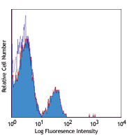
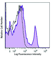
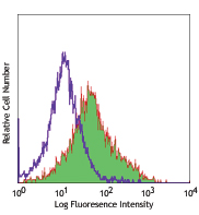
HEK293 cells were either untreated (negative control, open histogram), or treated with Oligomycin (positive control, filled histogram). The cells were fixed and permeabilized using the True-Phos™ Perm Buffer Set (Cat. No. 425401) and intracellularly stained with Purified anti-AMPKα Phospho (Thr172) (clone A20017A) or Purified Mouse IgG1, κ Isotype Control (open histogram, dashed line) (representative histogram for either untreated or treated cells) (Cat. No. 401402) followed by Alexa Fluor® 647 Goat anti-mouse IgG (Cat. No. 405322). -

HC staining of Purified anti-AMPKα Phospho (Thr172) on formalin-fixed paraffin-embedded human colon was performed following antigen retrieval using Tris-EDTA pH 9.0 Antigen Retrieval Buffer (Cat. No. 422703). Tissue sections were incubated with Alexa Fluor® 647 Goat anti-mouse only (Cat. No. 405322) (panel A) or with Purified anti-AMPKα Phospho (Thr172) (clone A20017A) followed by Alexa Fluor® 647 Goat anti-mouse (Cat. No. 405322) (panel B). Nuclei were counterstained with DAPI (Cat. No. 422801). Scale bar: 50 µm
| Cat # | Size | Price | Quantity Check Availability | Save | ||
|---|---|---|---|---|---|---|
| 600651 | 25 µg | 113€ | ||||
| 600652 | 100 µg | 264€ | ||||
AMP-activated protein kinase alpha (AMPKα) is one subunit of the αβγ heterotrimeric protein complex AMPK. It is a key regulator of metabolism and energy homeostasis in eukaryotes through induction of catabolic pathways and deactivation of anabolic. As an inhibitor of mammalian target of rapamycin complex 1 (mTORC1) pathway, AMPKα is involved in the regulation of cellular growth, cell cycle progression, autophagy, and may play dual roles as both tumor suppressor and promotor. AMPKα is activated via phosphorylation of Threonine residue 172 (Thr172) within the activation loop of the α subunit and is promoted by the allosteric binding of AMP and/or ADP to AMPK’s γ subunit. This phosphorylation is mediated by the serine/threonine kinase liver kinase B1 (LKB1) in conjunction with accessory subunits STRAD and MO25 and also by calcium/calmodulin-dependent protein kinase kinase 2 (CAMKK2 aka CAMKKβ). Activation is induced by increases in cellular AMP:ATP and ADP:ATP ratios following a decline in ATP levels as well as by cellular stress, DNA damage, glucose starvation, and fluxes in calcium and nutrient levels. Localization of AMPKα Thr172 at the spindle poles suggests a novel role for AMPK in mitotic spindle orientation. AMPKα is a key therapeutic target in the treatment of metabolic disorders and an emerging potential treatment target for cancer.
Product DetailsProduct Details
- Verified Reactivity
- Human, Mouse
- Antibody Type
- Monoclonal
- Host Species
- Mouse
- Immunogen
- Synthetic peptide of human AMPK alpha phosphorylated at Thr172
- Formulation
- Phosphate-buffered solution, pH 7.2, containing 0.09% sodium azide
- Preparation
- The antibody was purified by affinity chromatography.
- Concentration
- 0.5 mg/mL
- Storage & Handling
- The antibody solution should be stored undiluted between 2°C and 8°C.
- Application
-
WB - Quality tested
ICC, ICFC, IHC-P - Verified - Recommended Usage
-
Each lot of this antibody is quality control tested by western blotting. For western blotting, the suggested use of this reagent is 0.125 - 1.0 µg/mL. For immunocytochemistry, a concentration of 5.0 μg/mL is recommended. For flow cytometric staining using our True-Phos™ Perm Buffer, the suggested use of this reagent is ≤ 0.5 µg per million cells in 100 µL volume. For immunohistochemistry on formalin-fixed paraffin-embedded tissue sections, a concentration range of 1 - 10 µg/mL is suggested. It is recommended that the reagent be titrated for optimal performance for each application.
- Application Notes
-
This clone was tested for ICC using serum-starved untreated HEK293 cells (negative control) and HEK293 cells treated with 5.0 µM oligomycin for 30 minutes (positive control). Staining was expected to localize to the nucleus and cytoplasm. Three fix/perm methods were used: methanol fixation or 4% PFA fixation followed by permeabilization with either methanol or Triton X-100. Both PFA fixation followed by methanol permeabilization and PFA fixation followed by Triton X-100 permeabilization produced a strong signal with the expected localization. Methanol-only fixation only produced a faint signal.
- RRID
-
AB_2904438 (BioLegend Cat. No. 600651)
AB_2904438 (BioLegend Cat. No. 600652)
Antigen Details
- Structure
- AMPKα is a 552 amino acid protein with a predicted molecular weight of 63 kD.
- Distribution
-
Heart, Skeletal muscle, Kidney / Nucleus and cytoplasm
- Function
- Serine/threonine-protein kinase, transcription regulation, Wnt signaling, metabolism
- Ligand/Receptor
- ATP-binding, magnesium, Metal-binding, Nucelotide-binding
- Biology Area
- Cell Biology, Signal Transduction
- Molecular Family
- Phospho-Proteins, Protein Kinases/Phosphatase
- Antigen References
-
- Mihaylova MM, et al. 2011. Nat Cell Biol 13:1016-23.
- Stein SC, et al. 2000. Biochem J. 345:437-43.
- Thaiparambil JT, et al. 2012. Mol Cell Biol. 16:3203-17.
- Vara-Ciruelos D, et al. 2019. Open Biol. 9:190099.
- Willows R, et al. 2017. Biochem J. 474:3059-3073.
- Gene ID
- 5563 View all products for this Gene ID 5562 View all products for this Gene ID
- UniProt
- View information about AMPKalpha Phospho Thr172 on UniProt.org

Related FAQs
Other Formats
View All AMPKα Phospho (Thr172) Reagents Request Custom Conjugation| Description | Clone | Applications |
|---|---|---|
| Purified anti-AMPKα Phospho (Thr172) | A20017A | WB,ICC,ICFC,IHC-P |
| Alexa Fluor® 488 anti-AMPKα Phospho (Thr172) | A20017A | ICFC |
| Alexa Fluor® 647 anti-AMPKα Phospho (Thr172) | A20017A | IHC-P,ICFC |
Customers Also Purchased
Compare Data Across All Formats
This data display is provided for general comparisons between formats.
Your actual data may vary due to variations in samples, target cells, instruments and their settings, staining conditions, and other factors.
If you need assistance with selecting the best format contact our expert technical support team.
-
Purified anti-AMPKα Phospho (Thr172)

Whole cell extracts (15 µg total protein) from serum-starved... 
Whole cell extracts (15 µg total protein) from untreated C2C... 
Serum-starved untreated HEK293 cells (negative control, pane... 
HEK293 cells were either untreated (negative control, open h... 
HC staining of Purified anti-AMPKα Phospho (Thr172) on forma... -
Alexa Fluor® 488 anti-AMPKα Phospho (Thr172)

HEK293 cells untreated (negative control, open histogram) or... -
Alexa Fluor® 647 anti-AMPKα Phospho (Thr172)

IHC staining with Alexa Fluor® 647 anti-AMPKα Phospho (Thr17... 
Serum starved HEK293 cells were either untreated (low expres...


Follow Us