 Login / Register


- Clone
- A18040J (See other available formats)
- Regulatory Status
- RUO
- Other Names
- Cofilin 1, CFL1, CFL
- Isotype
- Mouse IgG1, κ
- Ave. Rating
- Submit a Review
- Product Citations
- publications

-

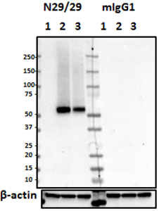
Western blot of purified anti-Cofilin 1 antibody (clone A18040J). Lane 1: Molecular weight marker; Lane 2: 20µg of mouse brain lysate; Lane 3: 40µg of rat brain lysate; Lane 4: 10µg of HeLa cell lysate; Lane 5: 10 µg of NIH3T3 cell lysate. The blots were incubated with 5 µg/mL of clone A8040J or mouse IgG overnight at 4°C, followed by incubation with HRP-labeled goat anti-mouse IgG (Cat. No. 405306). Enhanced chemiluminescence was used as the detection system. -

IHC staining of purified anti-Cofilin 1 antibody (clone A18040J) on formalin-fixed paraffin-embedded mouse colon tissue. Following antigen retrieval using Sodium Citrate H.I.E.R., the tissue was incubated with 5 µg/ml of the primary antibody overnight at 4°C. BioLegend's Ultra Streptavidin (USA) HRP Detection Kit (Multi-Species, DAB, Cat. No. 929901) was used for detection followed by hematoxylin counterstaining, according to the protocol provided. The image was captured with a 40X objective. Scale Bar: 50 µm -

IHC staining of purified anti-Cofilin 1 antibody (clone A18040J) on formalin-fixed paraffin-embedded rat colon tissue. Following antigen retrieval using Sodium Citrate H.I.E.R., the tissue was incubated with 1 µg/ml of the primary antibody overnight at 4°C. BioLegend's Ultra Streptavidin (USA) HRP Detection Kit (Multi-Species, DAB, Cat. No. 929901) was used for detection followed by hematoxylin counterstaining, according to the protocol provided. The image was captured with a 40X objective. Scale Bar: 50 µm -

ICC staining of purified anti-Cofilin 1 antibody (clone A18040J) on HeLa Cells. The cells were fixed with 4% PFA, permeabilized with a buffer containing 0.1% Triton X-100 and 0.25% BSA, and blocked with 2% normal goat serum and 0.02% BSA. The cells were then incubated with 2 µg/mL of the primary antibody overnight at 4°C, followed by incubation with 2.5 µg/mL of Alexa Fluor® 594 goat anti-mouse IgG for one hour at room temperature. The cells were co-stained with Flash Phalloidin™ Green 488 (Cat. No. 424201). The slide was mounted with fluoromount G with DAPI. The image was captured with a 60X objective. Scale bar: 20 µm
| Cat # | Size | Price | Quantity Check Availability | Save | ||
|---|---|---|---|---|---|---|
| 865301 | 25 µg | 85€ | ||||
| 865302 | 100 µg | 221€ | ||||
Cofilin 1 is a ubiquitously expressed actin-regulating protein that binds to and depolymerizes filamentous actin (F-actin), as well as inhibits G-actin polymerization in a pH-dependent mechanism. Cofilin 1 plays a major role in cell migration, particularly tumor cell migration and invasion. A recent study using colorectal cancer as model, compared expression of cofilin 1 in cancer tissue and adjacent non-tumor tissue and suggested that it may be used as a novel diagnostic tumor marker in various cancers4.
Product DetailsProduct Details
- Verified Reactivity
- Human, Mouse, Rat
- Antibody Type
- Monoclonal
- Host Species
- Mouse
- Immunogen
- Human CFL1 recombinant protein (a.a. Met 1 - Leu 166)
- Formulation
- Phosphate-buffered solution, pH 7.2, containing 0.09% sodium azide.
- Preparation
- The antibody was purified by affinity chromatography.
- Concentration
- 0.5 mg/mL
- Storage & Handling
- The antibody solution should be stored undiluted between 2°C and 8°C.
- Application
-
WB - Quality tested
IHC-P, ICC - Verified - Recommended Usage
-
Each lot of this antibody is quality control tested by Western blotting. For Western blotting, the suggested use of this reagent is 2.0 - 10 µg per mL. For immunohistochemistry on formalin-fixed paraffin-embedded tissue sections, a concentration range of 1.0 - 5.0 µg/mL is suggested. For immunocytochemistry, a concentration range of 2.0 - 10 μg/mL is recommended. It is recommended that the reagent be titrated for optimal performance for each application.
- Application Notes
-
For WB on brain lysates, 5 - 10 µg/mL is recommended.
- RRID
-
AB_2810771 (BioLegend Cat. No. 865301)
AB_2810772 (BioLegend Cat. No. 865302)
Antigen Details
- Structure
- Cofilin 1 is a 166 amino acid protein with an apparent molecular mass of ~19 kD.
- Distribution
-
Tissue Distribution: Immune system, gastrointestinal tract, lung, testis, and skin
Cellular Distribution: Nucleus, cytosol, plasma membrane, cytoskeleton, and mitochondrion - Interaction
- Cofilin 1 plays a role in cytoskeleton dynamics by regulating F-actin depolimerization
- Ligand/Receptor
- HSPH1, LIMK1, F-actin
- Cell Type
- Astrocytes, Dopaminergic Neurons, GABAergic Neurons, Glutamatergic Neurons, Mature Neurons, Microglia, Neural Stem Cells, Neurons, Oligodendrocytes
- Biology Area
- Cancer Biomarkers, Cell Biology, Cell Motility/Cytoskeleton/Structure, Cell Proliferation and Viability, Cell Structure, Neurodegeneration, Neuroscience, Synaptic Biology
- Molecular Family
- Cytoskeletal Proteins, GTPases, Scaffold Proteins
- Antigen References
-
- Kanellos G and Frame MC. 2016. J Cell Sci. 129(17):3211
- Walsh KP, et al. 2014. Prion. 8(6):375
- Xu P, et al. 2018. Front Plant Sci. 9:1528
- Mousavi S, et al. 2018. J Gastrointest Oncol. 9(5):791
- Gene ID
- 1072 View all products for this Gene ID 12631 View all products for this Gene ID 29271 View all products for this Gene ID
- UniProt
- View information about Cofilin 1 on UniProt.org

Related FAQs
Other Formats
View All Cofilin 1 Reagents Request Custom Conjugation| Description | Clone | Applications |
|---|---|---|
| Purified anti-Cofilin 1 | A18040J | WB,IHC-P,ICC |
Customers Also Purchased
Compare Data Across All Formats
This data display is provided for general comparisons between formats.
Your actual data may vary due to variations in samples, target cells, instruments and their settings, staining conditions, and other factors.
If you need assistance with selecting the best format contact our expert technical support team.


Follow Us