 Login / Register


- Clone
- A16009C (See other available formats)
- Regulatory Status
- RUO
- Other Names
- 40S ribosomal protein S6, Ribosomal Protein S6
- Isotype
- Mouse IgG2b, κ
- Ave. Rating
- Submit a Review
- Product Citations
- publications

-

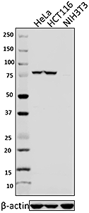
Total cell lysates (15 µg protein) from HeLa, Jurkat and NIH3T3 were resolved by 4-20% Tris-glycine gel electrophoresis, transferred to nitrocellulose, and probed with 0.25 µg/mL purified anti-RPS6 (clone A16009C) antibody. Proteins were visualized using a goat anti-mouse-IgG secondary antibody conjugated to HRP and chemiluminescence detection. Direct-Blot™ HRP anti-β-actin (clone 2F1-1) antibody was used as a loading control. -

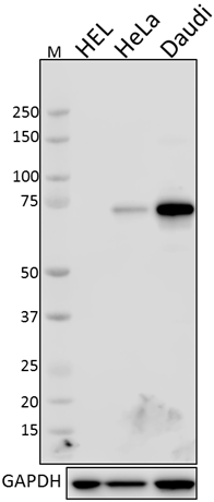
Total lysates (15 µg protein) from 293T and 293T/RPS6 CRISPR/Cas9 knockdown (KD) cells were resolved by electrophoresis (4-20% Tris-Glycine gel), transferred to nitrocellulose, and probed with 1:1000 diluted (0.5 µg/mL) purified anti-RPS6 antibody, clone A16009C (upper). Proteins were visualized using chemiluminescence detection by incubating with 1:3000 dilution of HRP goat anti-mouse-IgG secondary antibody (Cat. No. 405306). 1:2000 dilution of Direct-Blot™ HRP anti-β-actin Antibody (Cat. No. 643807) was used as a loading control (lower). Lane M: MW ladder. -

293T (human embryonic kidney cell line) fixed with Fixation Buffer (Cat No. 420801), permeabilized with 10X Permeabilization Buffer (Cat No. 421002), and intracellularly stained with purified anti-RPS6 (clone A16009C) (filled histogram) or purified Mouse IgG2b, κ isotype control (open histogram), followed by anti-mouse IgG PE. -

IHC staining of purified anti-RPS6 antibody (clone A16009C) on formalin-fixed paraffin-embedded human thymus tissue. Following antigen retrieval using 10 mM Tris-base, 1 mM EDTA in deionized water, pH 9.0, the tissue was incubated with 5 µg/mL of the primary antibody overnight at 4°C, followed by incubation with 2.5 µg/mL of Alexa Fluor® 647 goat anti-mouse IgG (red) (Cat. No. 405322) for one hour at room temperature. Nuclei were counterstained with DAPI (blue), and the slide was mounted with ProLong™ Gold Antifade Mountant. The image was captured with a 20x objective. Scale bar: 50 µm
| Cat # | Size | Price | Quantity Check Availability | Save | ||
|---|---|---|---|---|---|---|
| 691802 | 100 µg | 203€ | ||||
The eukaryotic ribosome is composed of 40S and 60S subunits, which is involved in the physiological process of protein translation. In many organisms, ribosomal proteins undergo various post-translational modifications, including phosphorylation, acetylation, methylation, O-linked b-N-acetylglucosaminylation, and ubiquitylation. Ribosomal proteinS6 (RPS6), the first identified ribosomal protein which can be phosphorylated, is a component of eukaryotic ribosomal 40S subunit. Functional studies of RPS6 phosphorylation suggested its role in the regulation of protein synthesis, cell proliferation, and glucose homeostasis. The phosphorylation of RPS6 can be triggered by multiple stimuli including growth factors, tumor promoting agents, and mitogens. The inhibition of RPS6 phosphorylation was found mainly under stress conditions such as hypoxia, heat shock, and hyperosmolarity. RPS6 phosphorylation is also commonly used as a marker for neuronal activity and a read out of mammalian target of rapamycin complex 1 (mTORC1) activity.
Product DetailsProduct Details
- Verified Reactivity
- Human, Mouse
- Antibody Type
- Monoclonal
- Host Species
- Mouse
- Immunogen
- Human S6 peptide.
- Formulation
- Phosphate-buffered solution, pH 7.2, containing 0.09% sodium azide.
- Preparation
- The antibody was purified by affinity chromatography.
- Concentration
- 0.5 mg/ml
- Storage & Handling
- The antibody solution should be stored undiluted between 2°C and 8°C.
- Application
-
WB - Quality tested
ICFC, KO/KD-WB, IHC-P - Verified - Recommended Usage
-
Each lot of this antibody is quality control tested by Western blotting. For Western blotting, the suggested use of this reagent is 0.1 - 1.0 µg per ml. For intracellular flow cytometric staining, the suggested use of this reagent is ≤ 0.25 µg per million cells in 100 µl volume. For immunohistochemistry on formalin-fixed paraffin-embedded tissue sections, a concentration range of 1.0 - 5.0 µg/ml is suggested.It is recommended that the reagent be titrated for optimal performance for each application.
- Product Citations
-
- RRID
-
AB_2629833 (BioLegend Cat. No. 691802)
Antigen Details
- Structure
- 249 amino acids with a predicted molecular weight of 28.7kD.
- Distribution
-
Cytoplasm and nucleus.
- Function
- Ribosomal protein S6 is the major substrate of protein kinases in eukaryote ribosomes. The phosphorylation is stimulated by growth factors, tumor promoting agents, and mitogens. It is dephosphorylated at growth arrest.
- Interaction
- NCBP1, PASK.
- Biology Area
- Cell Biology, Signal Transduction
- Antigen References
-
1. Kressler A, et al. 2010. Biochim. Biophys. Acta. 1803:673.
2. Biever A, et al. 2015. Front. Mol. Neurosci. 8:75.
3. Ventimiglia FA, Wool IG. 1974. Proc. Natl. Acad. Sci. USA 71:350.
4. Lastick SM, et al. 1977. Mol. Gen. Genet. 152:223.
5. Ruvinsky I, et al. 2005. Genes Dev. 19:2199.
6. Meyuhas O. 2008. Int. Rev. Cell. Mol. Biol. 268:1.
7. Meyuhas O. 2015. Int. Rev. Cell. Mol. Biol. 320:41. - Gene ID
- NA
- UniProt
- View information about RPS6 on UniProt.org

Related FAQs
Other Formats
View All Reagents Request Custom Conjugation| Description | Clone | Applications |
|---|---|---|
| Purified anti-RPS6 | A16009C | WB,ICFC,KO/KD-WB,IHC-P |
| PE anti-RPS6 | A16009C | ICFC |
Customers Also Purchased
Compare Data Across All Formats
This data display is provided for general comparisons between formats.
Your actual data may vary due to variations in samples, target cells, instruments and their settings, staining conditions, and other factors.
If you need assistance with selecting the best format contact our expert technical support team.
-
Purified anti-RPS6

Total cell lysates (15 µg protein) from HeLa, Jurkat and NIH... 
Total lysates (15 µg protein) from 293T and 293T/RPS6 CRISPR... 
293T (human embryonic kidney cell line) fixed with Fixation ... 
IHC staining of purified anti-RPS6 antibody (clone A16009C) ... -
PE anti-RPS6

HEK 293T cells were fixed, permeabilized, and intracellularl...


Follow Us