
 Login / Register


- Clone
- TUJ1 (See other available formats)
- Regulatory Status
- RUO
- Other Names
- CDCBM, CDCBM1, CFEOM3, CFEOM3A, FEOM3, TUBB4, Tubulin beta-3 chain, tubulin beta-III, tubulin beta-4 chain, class III beta-tubulin
- Previously
-
Covance Catalog# MMS-435P
- Isotype
- Mouse IgG2a, κ
- Ave. Rating
- Submit a Review
- Product Citations
- 818 publications


| Cat # | Size | Price | Quantity Check Availability | Save | ||
|---|---|---|---|---|---|---|
| 801213 | 25 µL | 85€ | ||||
| 801201 | 100 µL | 212€ | ||||
| 801202 | 250 µL | 313€ | ||||
Tubulin is the main component of microtubules. In adults, tubulin beta 3 (TUBB3) is primarily expressed in neurons and is commonly used as a neuronal marker. It plays an important role in neuronal cell proliferation and differentiation. Mutations in this gene cause congenital fibrosis of the type 3 extraocular muscles. Tubulin beta 3 (TUBB3) is also found in a wide range of tumors. Studies indicate that it is a predictive and prognostic marker in various tumors.
Product DetailsProduct Details
- Verified Reactivity
- Human, Mouse, Rat
- Antibody Type
- Monoclonal
- Host Species
- Mouse
- Immunogen
- This antibody was raised against microtubules derived from rat brain.
- Formulation
- Phosphate-buffered solution + 0.03% Thimerosal.
- Preparation
- The antibody was purified by affinity chromatography.
- Concentration
- 1.0 mg/ml
- Storage & Handling
- The antibody solution should be stored undiluted between 2°C and 8°C. Please note the storage condition for this antibody has been changed from -20°C to between 2°C and 8°C. You can also check your vial or your CoA to find the most accurate storage condition for this antibody.
- Application
-
IHC-P - Quality tested
WB, ICC - Verified
FC - Reported in the literature, not verified in house
SB - Community verified - Recommended Usage
-
Each lot of this antibody is quality control tested by formalin-fixed paraffin-embedded immunohistochemical staining. For immunohistochemistry, a concentration range of 1.0 - 5.0 µg/ml is suggested. For Western blotting, the suggested use of this reagent is 1.0 - 5.0 µg/ml. For immunocytochemistry, a concentration range of 1.0 - 5.0 μg/ml is recommended. It is recommended that the reagent be titrated for optimal performance for each application.
- Application Notes
-
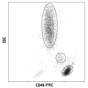
Additional reported applications (for the relevant formats) include: flow cytometry4, immunofluorescence microscopy1-5,7, immunohistochemistry5,7, Western blotting8, and spatial biology (IBEX)9,10.
This antibody is well characterized and highly reactive to neuron specific Class III ß-tubulin (ßIII). TUJ1 does not identify ß-tubulin found in glial cells. TUJ1 recognizes an epitope located within the last 15 C-terminal residues8. - Additional Product Notes
-
This product has been verified for IHC-F (Immunohistochemistry - frozen tissue sections) on the NanoString GeoMx® Digital Spatial Profiler. The GeoMx® enables researchers to perform spatial analysis of protein and RNA targets in FFPE and fresh frozen human and mouse samples. For more information about our spatial biology products and the GeoMx® platform, please visit our spatial biology page.
-
Application References
(PubMed link indicates BioLegend citation) -
- Nishimura K, et al. 2017. PLoS One. 12(1): e0170568. (ICC)
- Jongbloets J, et al. 2017. Nat Commun. 8: 14666. (ICC) PubMed
- Liu W.J, et al. 2015. Eur J Histochem. 59(1): 2464. (ICC) PubMed
- Chintalapudi SR, et al. 2016. Front Aging Neursci. 8:93. (FC, ICC) PubMed
- Ambasudhan R, et al. 2011. Cell Stem Cell. 9(2):113. (IHC, ICC)
- Hu X., et al. 2006. Nature Neuroscicene. 9(12):1520. (WB) PubMed
- Zechner D., et al. 2003. Develop Biology. 258(2):406. (ICC, IHC)
- Lee MK, et al. 1990. Proc. Natl. Acad. Sci. USA 18:7195. (WB)
- Radtke AJ, et al. 2020. Proc Natl Acad Sci USA. 117:33455-33465. (SB) PubMed
- Radtke AJ, et al. 2022. Nat Protoc. 17:378-401. (SB) PubMed
- Product Citations
-
- RRID
-
AB_2313773 (BioLegend Cat. No. 801213)
AB_2313773 (BioLegend Cat. No. 801201)
AB_2313773 (BioLegend Cat. No. 801202)
- Disclaimer
-
Covered by US patents 5,675,063 and 7,429,487. Sold under license from Epitomics.
Antigen Details
- Structure
- Tubulin β 3 is a 450 amino acid protein with a molecular mass of ~50 kD.
- Distribution
-
Tissue distribution: central and peripheral nervous system.
Cellular distribution: cytosol, cytoskeleton and nucleus. - Function
- Tubulin β 3 is the major constituent of microtubules, and plays a critical role in proper axon guidance and maintenance.
- Interaction
- Alpha tubulin, kinesin and dynein.
- Cell Type
- Mature Neurons, Neural Stem Cells
- Biology Area
- Cell Biology, Neuroscience, Neuroscience Cell Markers, Stem Cells
- Molecular Family
- Microtubules
- Antigen References
-
1. Zhao X, et al. 2017. Med Sci Monit. 22: 3915.
2. Lebok P, et al. 2016. Oncol Lett. 11(3):1987.
3. Du J, et al. 2015. BMC Cancer. 15:536. PubMed
4. Rogue DM., et al. 2013. Clin Exp Metastasis. 31(1): 101.
5. Ploussard G, et al. 2010. Cancer Res. 70(22):9253. PubMed - Gene ID
- 10381 View all products for this Gene ID
- UniProt
- View information about Tubulin beta-3 on UniProt.org

Related FAQs
- If an antibody clone has been previously successfully used in IBEX in one fluorescent format, will other antibody formats work as well?
-
It’s likely that other fluorophore conjugates to the same antibody clone will also be compatible with IBEX using the same sample fixation procedure. Ultimately a directly conjugated antibody’s utility in fluorescent imaging and IBEX may be specific to the sample and microscope being used in the experiment. Some antibody clone conjugates may perform better than others due to performance differences in non-specific binding, fluorophore brightness, and other biochemical properties unique to that conjugate.
- Will antibodies my lab is already using for fluorescent or chromogenic IHC work in IBEX?
-
Fundamentally, IBEX as a technique that works much in the same way as single antibody panels or single marker IF/IHC. If you’re already successfully using an antibody clone on a sample of interest, it is likely that clone will have utility in IBEX. It is expected some optimization and testing of different antibody fluorophore conjugates will be required to find a suitable format; however, legacy microscopy techniques like chromogenic IHC on fixed or frozen tissue is an excellent place to start looking for useful antibodies.
- Are other fluorophores compatible with IBEX?
-
Over 18 fluorescent formats have been screened for use in IBEX, however, it is likely that other fluorophores are able to be rapidly bleached in IBEX. If a fluorophore format is already suitable for your imaging platform it can be tested for compatibility in IBEX.
- The same antibody works in one tissue type but not another. What is happening?
-
Differences in tissue properties may impact both the ability of an antibody to bind its target specifically and impact the ability of a specific fluorophore conjugate to overcome the background fluorescent signal in a given tissue. Secondary stains, as well as testing multiple fluorescent conjugates of the same clone, may help to troubleshoot challenging targets or tissues. Using a reference control tissue may also give confidence in the specificity of your staining.
- How can I be sure the staining I’m seeing in my tissue is real?
-
In general, best practices for validating an antibody in traditional chromogenic or fluorescent IHC are applicable to IBEX. Please reference the Nature Methods review on antibody based multiplexed imaging for resources on validating antibodies for IBEX.
Other Formats
View All Tubulin β 3 (TUBB3) Reagents Request Custom Conjugation| Description | Clone | Applications |
|---|---|---|
| Alexa Fluor® 488 anti-Tubulin β 3 (TUBB3) | TUJ1 | ICC,IHC-P,ICFC,3D IHC,SB |
| Purified anti-Tubulin β 3 (TUBB3) | TUJ1 | IHC-P,WB,ICC,FC,SB |
| Alexa Fluor® 594 anti-Tubulin β 3 (TUBB3) | TUJ1 | ICC,3D IHC,IHC-P |
| Alexa Fluor® 647 anti-Tubulin β 3 (TUBB3) | TUJ1 | ICC,ICFC,3D IHC,IHC-P |
| HRP anti-Tubulin β 3 (TUBB3) | TUJ1 | WB,IHC-P |
| Biotin anti-Tubulin β 3 (TUBB3) | TUJ1 | WB,IHC-P |
| APC anti-Tubulin β 3 (TUBB3) | TUJ1 | ICFC |
| PE/Cyanine7 anti-Tubulin β 3 (TUBB3) | TUJ1 | ICFC |
| PerCP/Cyanine5.5 anti-Tubulin β 3 (TUBB3) | TUJ1 | ICFC |
| PE anti-Tubulin β 3 (TUBB3) | TUJ1 | ICFC |
Customers Also Purchased
Compare Data Across All Formats
This data display is provided for general comparisons between formats.
Your actual data may vary due to variations in samples, target cells, instruments and their settings, staining conditions, and other factors.
If you need assistance with selecting the best format contact our expert technical support team.
-
Alexa Fluor® 488 anti-Tubulin β 3 (TUBB3)

CHO cell line expressing Tubulin β 3 stained with Alexa Fluo... 
IHC staining of Alexa Fluor® 488 anti-Tubulin β 3 (TUBB3) an... 
IHC staining of Alexa Fluor® 488 anti-Tubulin β 3 (TUBB3) an... 
Human lung adenocarcinoma cell line A549 was treated with Fi... 
Paraformaldehyde-fixed (4%), 500 µm-thick mouse kidney tissu... 
Confocal image of human jejunum sample acquired using the IB... -
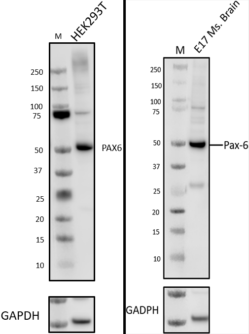
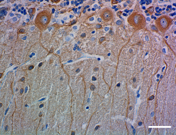
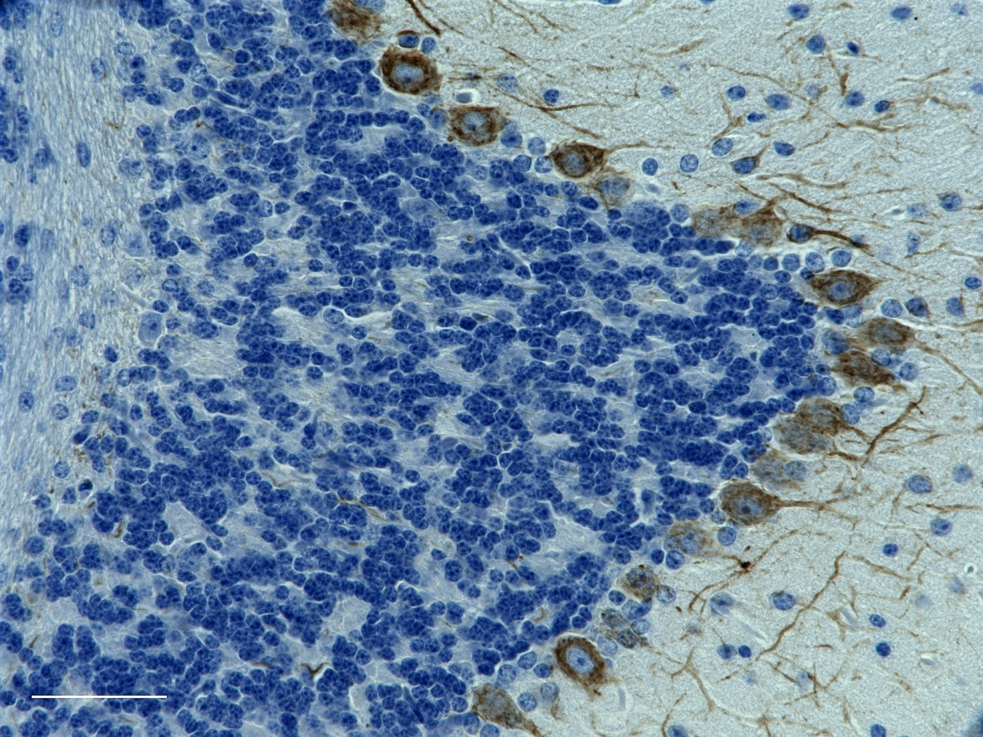
Purified anti-Tubulin β 3 (TUBB3)

IHC staining of purified anti-Tubulin β 3 (TUBB3) antibody (... 
IHC staining of purified anti-Tubulin β 3 (TUBB3) antibody (... 
Western blot of purified anti-Tubulin β 3 (TUBB3) antibody (... 
ICC staining of purified anti-Tubulin β 3 (TUBB3) antibody (... 
Methanol fixed superior cervical ganglion neurons were stain... -
Alexa Fluor® 594 anti-Tubulin β 3 (TUBB3)

ICC staining of Alexa Fluor® 594 anti-Tubulin β 3 (TUBB3) (c... 
Paraformaldehyde-fixed (4%), 500 μm-thick mouse kidney secti... 
A: IHC staining of Alexa Fluor® 594 anti-Tubulin β 3 (TUBB3)... -
Alexa Fluor® 647 anti-Tubulin β 3 (TUBB3)

ICC staining of Alexa Fluor® 647 anti-Tubulin β 3 (TUBB3) an... 
ICC staining of Alexa Fluor® 647 anti-Tubulin β 3 (TUBB3) (c... 
Paraformaldehyde-fixed (4%), 500 μm-thick mouse kidney secti... 
IHC staining of Alexa Fluor® 647 anti-Tubulin β 3 (TUBB3) (c... -
HRP anti-Tubulin β 3 (TUBB3)

IHC staining of HRP anti-Tubulin β 3 (TUBB3) antibody (clone... 
IHC staining of anti-Tubulin β 3 (TUBB3) antibody (clone TUJ... 
Western blot of HRP anti-Tubulin β 3 (TUBB3) antibody (clone... -
Biotin anti-Tubulin β 3 (TUBB3)

Western blot of Biotin anti-Tubulin β 3 (TUBB3) antibody (cl... 
IHC staining of Biotin anti-Tubulin β 3 (TUBB3) antibody (cl... -
APC anti-Tubulin β 3 (TUBB3)

Human lung adenocarcinoma cell line A549 was treated with Fi... -
PE/Cyanine7 anti-Tubulin β 3 (TUBB3)

Human lung adenocarcinoma cell line A549 was treated with Fi... -
PerCP/Cyanine5.5 anti-Tubulin β 3 (TUBB3)

Human lung adenocarcinoma cell line A549 was treated with Fi... -
PE anti-Tubulin β 3 (TUBB3)

Human lung adenocarcinoma cell line A549 was treated with Fi...


Follow Us