Intermediate Filaments


Intermediate filaments (IFs) are structural components of the cytoskeleton that make up polymers with an average diameter of 10 nm. IFs are categorized into six different types (I-VI). Most IFs are cytosolic except type V nuclear lamins which are localized to the nucleus. IFs provide mechanical support, help maintain cellular shape and rigidity, and anchor organelles, such as the nucleus and desmosomes, in place. Type V IFs are involved in the formation of the nuclear lamina, a meshwork composed of lamins and lamin-associated proteins. It lines the inner nuclear membrane and helps govern the shape and organization of the nucleus.


The expression and composition of intermediate filaments can be species- and tissue-dependent in vertebrates. The table below summarizes type I to VI IFs:


Type I: Acidic keratins

Type IV: Neurofilaments

Type II: Basic keratins

Type V: Lamins

Type III: GFAP, Vimentin

Type VI: Nestin


Type I and II IFs: Keratins


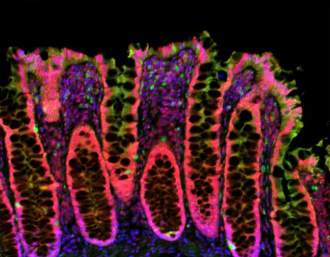
Keratins, also known as cytokeratins (CKs), are intracytoplasmic cytoskeletal proteins found in epithelial tissue that provide resistance to mechanical stress. Keratins are subdivided into type I (acidic) and type II (basic) keratins, and further numbered within each category. This classification is done in order of decreasing size from high to low molecular weight. Keratins 9-23 and 1-8 make up the acidic and basic keratins, respectively. They are expressed in a tissue- and differentiation state-specific manner and form obligate, non-covalent heteropolymers between type 1 and type 2 keratins. For instance, keratins 8 and 18 are the primary keratin pair in hepatocytes, keratins 7 and 19 are expressed in intestinal cells, and keratins 5 and 14 exist in keratinocytes of stratified squamous epithelia1.


The use of pan- or broad-spectrum cytokeratin antibodies in immunohistochemistry (IHC) has been a reliable tissue-based application for the detection and identification of the origin of various human tumors and can distinguish different types of tumors. In addition to offering antibodies for specific keratins, we carry well-known pan CK antibodies, including clones AE-1, AE-3, AE-1/AE-3, C-11, and OSCAR.  The reactivity of each of these clones is listed in the table below:

Clone

Type 2 (basic)

Type 1 (acidic)


1

2

3

4

5

6

7

8

9

10

11

12

13

14

15

16

17

18

19

20

AE-1


x


x

x

x


x


AE-3

x

x

x

x

x

x

x

x


AE-1/AE-3

x

x

x

x

x

x

x

x


x


x

x

x


x


C-11


x

x

x


x


x


x


x


OSCAR


x

x


x

x


Type III IFs: Vimentin


Vimentin is a major IF cytoskeletal component that is primarily expressed in cells of mesenchymal origin. These include fibroblasts, endothelial cells, and immune cells like macrophages and neutrophils. Vimentin is responsible for maintaining cell shape and anchoring organelles such as the nucleus, endoplasmic reticulum (ER), and mitochondria, in place. Vimentin is predominantly a cytosolic protein, however, it can also be secreted to the extracellular space or become localized at the cell surface. Assembly and disassembly of intracellular vimentin networks is a dynamic process that is driven by post-translational modifications (PTMs) such as phosphorylation2.


In addition to stabilizing cytoskeletal interactions, vimentin plays a role in apoptosis, aggresome formation, and immune system activation. For example, PTMs are implicated in cell surface localization of vimentin, where it can act as a receptor for viruses, and induce the release of proinflammatory cytokines that can trigger tissue damage in response to virus infection2.


Because of its high level and constitutive expression in mesenchymal cells, vimentin serves as a good marker to differentiate mesenchymal from epithelial cells. It can also be used to identify cells that are undergoing an epithelial-to-mesenchymal transition (EMT) during development and in the early stages of cancer cell metastasis.


We provide antibodies for the detection of vimentin in western blotting and microscopy applications. Our directly-conjugated formats provide ease of co-staining with other markers to distinguish different cells, organelles, and cancer types.


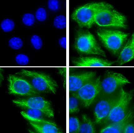
Immunofluorescence of A431 cells with Chicken IgG Y Isotype control (Negative) or Vimentin primary antibody. Alexa Fluor® 488 (Green) Goat anti-Chicken IgG was used as secondary antibody. Nuclei were counterstained with DAPI (Blue).

Type IV IFs: Neurofilaments


Neurofilaments (NFs) are the major components of the neuronal cytoskeleton and are essential for providing structural support and maintenance of axon caliber. Three mammalian neurofilament subunits, NF-L, NF-M, and NF-H, are classified based on their molecular weights. Filament assembly, function, and molecular interactions of neurofilaments can be regulated by phosphorylation. Aberrant phosphorylation of NF-M and NF-H leads to their accumulation in neuronal cell bodies in Alzheimer’s disease and disruption of NF transport to axons. NFs can also be released from damaged or diseased neurons into blood or CSF. Elevated levels of NFs in the serum or CSF can serve as a biomarker for neuronal injury or degeneration3.


Antibodies against NFs are an ideal tool for distinguishing neurons from glial cells, which do not express NFs. Furthermore, antibodies that can detect NF modifications by immunostaining are of great diagnostic value for neuropathology detection, since abnormal NF modifications, such as hyperphosphorylation, have been associated with neurodegenerative diseases.


Detection of neurofilaments allows for the visualization of neuronal axons, dendrites, and cell bodies depending on the clone utilized. We’ve created a large collection of purified and conjugated antibody formats and a sampler kit for both native and modified forms of NFs. Our selection of Alexa Fluor® conjugates enables multiplexed immunofluorescence staining. Examples of clones reactivities are included in the table below:


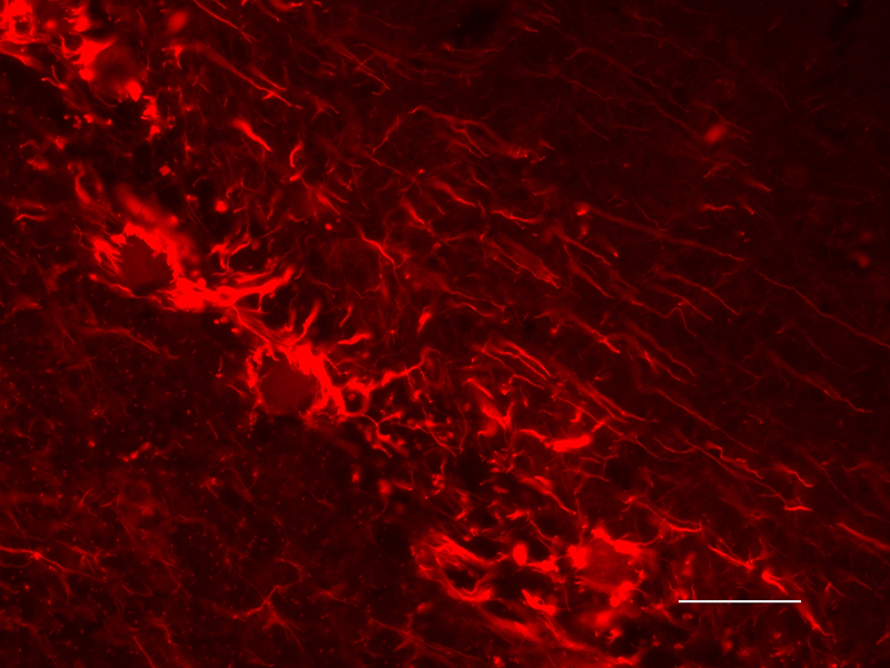
Formalin-fixed paraffin-embedded human brain tissue stained with purified anti-Neurofilament L (NF-L) antibody (clone NFL3) and Alexa Fluor® 594 Goat anti-Mouse IgG (red) used as secondary antibody.

Specificity

Clone

Neuronal Components


Axon

Dendrite

Cell Body

NF-L

NFL3

x


x

NF-H (phospho)

SMI 31

x


NF-H

SMI 32

x

x

x

NF-H/NF-M (phospho)

SMI 35

x


Type V IFs: Lamins


Lamins are nuclear IFs that assemble into filamentous structures to form a scaffold required for maintaining the nuclear structure. They also help anchor the nucleus to the ER. Lamins are predominantly involved in forming the nuclear lamina, however, a small fraction is also found localized to the nucleoplasm. Lamins are divided into two classes: A-type and B-type. The A-type lamins are encoded by the LMNA gene and can produce two isoforms, lamin A and lamin C, by alternative splicing. B-type lamins are also present in multiple isoforms. Lamin B1 and lamin B2 are the most common and are encoded by LMNB1 and LMNB2 genes, respectively. The expression of A-type lamins is developmentally regulated, and their expression typically occurs at later stages of development or after birth. B-type lamins are expressed in all cell types throughout development.


Lamins are involved in many nuclear processes including gene expression by organizing the chromatin into active and inactive domains. Because of their close association with the nuclear envelope and chromatin, lamins are also targets for apoptosis-induced nuclear deformation. Activation of p53 in response to various apoptotic stimuli can cause nuclear deformation by binding to and stabilizing lamin A/C4. Lamins can associate with several transcription factors and influence transcription. For instance, lamin B1 can associate with Oct-1 and play an essential role in transcriptional regulation of the cellular oxidative stress response genes5. Mutations in lamin genes result in disorders that are collectively termed laminopathies. These mutations can affect muscle, bone, and nerve cells, and includes disorders that range from muscular dystrophy to Hutchinson–Gilford progeria6. Utilize antibodies against lamins to detect the expression levels or localization of these proteins, or to assess nuclear integrity.

Type VI IFs: Nestin


Nestin interacts with other IF proteins including vimentin, desmin, and internexin to form heterodimers and mixed polymers. Nestin is expressed in neural stem and progenitor cells, and early stages of development in the central nervous system (CNS). Upon differentiation, and during neuro- and gliogenesis, nestin is replaced by cell type-specific intermediate filaments such as neurofilaments and glial fibrillary acidic protein (GFAP). Nestin distribution and expression patterns in proliferating cells suggest that it participates in the structural organization of the cell by regulating the assembly and disassembly of intermediate filaments. The function of nestin itself is highly regulated by phosphorylation. Nestin expression is induced in adults under pathological conditions such as glial scar formation after CNS injury7. Antibodies against nestin can be used as a predominant marker to describe stem and neural progenitor cells in the mammalian CNS.

References

  1. Karantza V. Keratins in health and cancer: more than mere epithelial cell markers. Oncogene 30: 127–138 (2011).
  2. Ramos I, et al. Vimentin as a Multifaceted Player and Potential Therapeutic Target in Viral Infections. Int. J. Mol. Sci. 21: 4675 (2020).
  3. Yyan A, et al. Neurofilaments and Neurofilament Proteins in Health and Disease. Cold Spring Harb Perspect Biol 9(4):1 (2017).
  4. Lindenboim L, et al. Apoptotic stress induces Bax-dependent, caspase-independent redistribution of LINC complex nesprins. Cell Death Discov. 6, 90 (2020). 
  5. Malhas AN, et al. Lamin B1 controls oxidative stress responses via Oct-1. J. Cell Biol. 184:1 45 (2009).
  6. Donnaloja F, et al. Lamin A/C Mechanotransduction in Laminopathies. Cells. 24;9(5):1306 (2020).
  7. Michalczyk K and Ziman M. Nestin structure and predicted function in cellular cytoskeletal organisation. Histol Histopathol. 20(2):665-71 (2005). 
ProductsHere

Login / Register
Remember me
Forgot your password? Reset password?
Create an Account